
Introduction of Auramine-Phenol stain
Table of Contents
Auramine-Phenol is a fluorochrome stain used to visualize acid-fast structures of various microorganisms, especially Mycobacterium tuberculosis and in modified form for Mycobacterium leprae, Nocardia species, Cryptosporidium parvum, Cyclospora cayetanensis , Isospora belli, and fungal spores. Ziehl-Neelsen (hot), and Kinyoun (cold) are still widely used methods to detect acid-fast structures in these organisms in developing countries but the sensitivity is high of fluorochrome stain. The acid fastness of Mycobacterium tuberculosis is due to having a thick cell wall composed of waxes and lipids that has a high content of mycolic acid.
Principle of Auramine-Phenol Stain
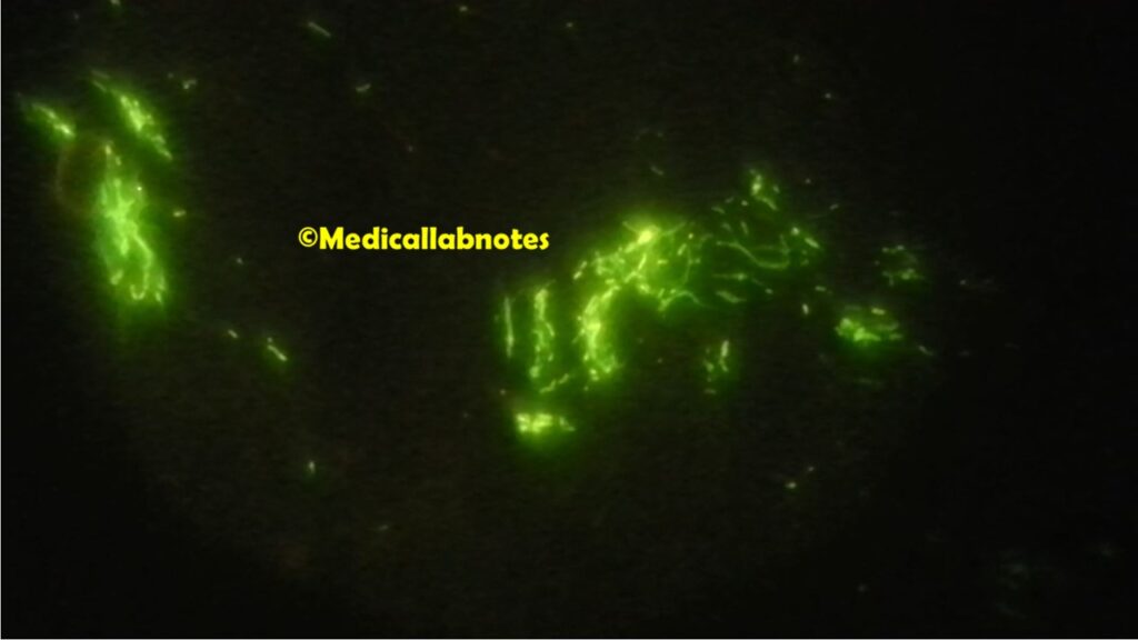
Auramine is the fluorochrome dye that forms a complex with mycolic acids found in the acid-fast cell wall of organisms that resist decolorization by acid-alcohol. Potassium permanganate, counterstain renders tissue and its debris nonfluorescent, therefore reducing the possibility of artifacts. The cellular structures visualized under U-V appear bright yellow or brilliant greenish-yellow against a dark background.
Test Requirements for Auramine-Phenol Stain
- Auramine-phenol stain (Primary Stain)
- 1 % Acid alcohol ( Decolorizer)
- 0.1% Potassium Permanganate
- Test specimen
- Slide ( clean and grease-free)
- Pencil ( diamond if possible)
- Slide racks
- Bunsen burner
- Inoculating loop or sterile bamboo stick
- Two positive slides, one for a heavy load and another for a weak load of AFB to maintain quality control
Staining Procedure of Auramine-Phenol Stain
- Smear preparation from sputum specimen
Take a purulent part of the sputum to a slide and make a thin smear using an inoculating loop or bamboo stick. Cover an area of approximately 2 cm square and spread the smear using circular movements. Allow it to air dry. Finally, perform heat-fixing passing the dried slide, smear facing upward, 2 to 3 times through the blue cone of a burner flame.
- Staining
- Put the fixed smear on a staining rack and flood the smear with auramine-phenol for 15 minutes. Do not let the smear dry.
- Wash off the stain with clean water.
- Decolorize the smear by covering it with acid-alcohol for 3-5 minutes. ( But in this case, weak acid-fast organisms like Nocradia, Cryptosporidium, etc use 0.1 % acid alcohol i.e. a modified form of auramine-phenol stain.)
- Wash off the acid alcohol with clean water.
- Now cover the smear with potassium permanganate for 15 seconds. Do not allow the smear to dry.
- Rinse thoroughly with distilled water and air dry.
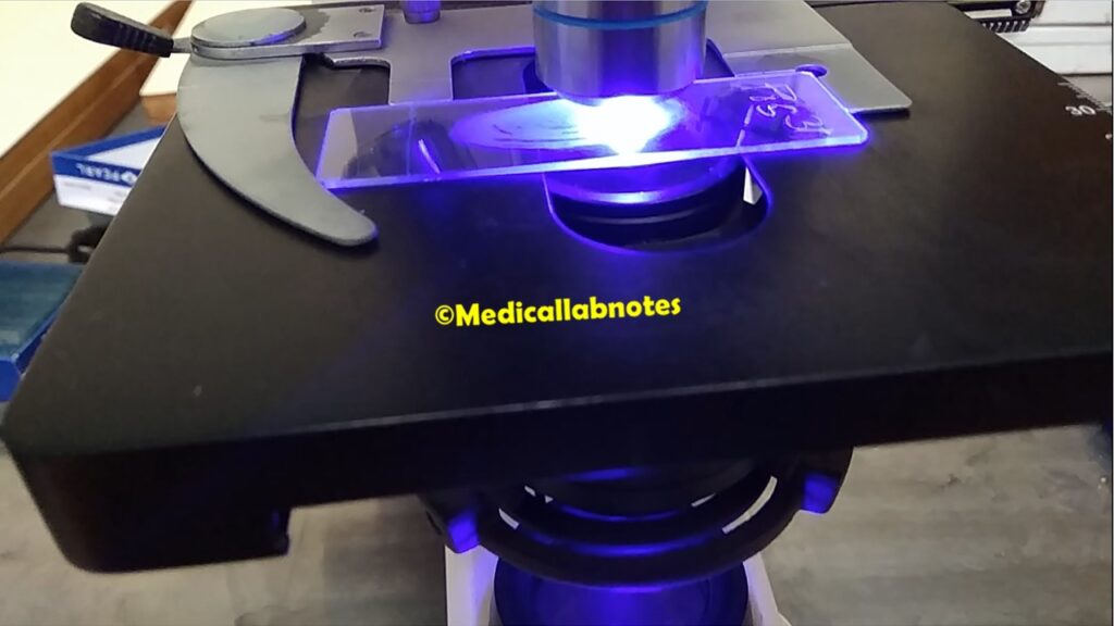
- Examine the smear with a fluorescence microscope and use the 10X objective to focus the smear. Finally, observe the smear using the 40 X objective for acid-fast structures or acid-fast bacilli (AFB).

Result and Interpretation of Auramine-Phenol Stain
Test Positive: Acid-fast organisms fluoresce bright yellow or reddish-orange against a dark background.
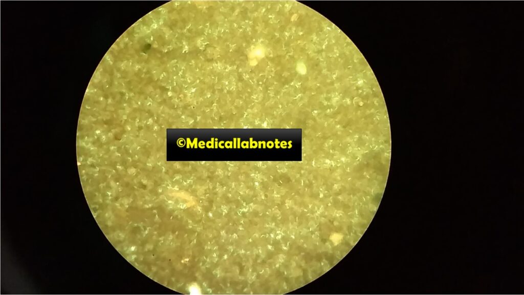
Negative Test: Non-acid-fast organisms will not fluoresce
Reporting Sputum Smear
According to IUATLD (International Union against Tuberculosis and Lung Diseases) and WHO guidelines-
Report using 40 x objective and 10 x eyepiece
Scanty AFB 1-19 AFB in 40 fields
+ 20-199 AFB in 40 fields
++ 5-50 AFB per field (examine 20 fields)
+++ >50 AFB per field ( examine 8 fields)
To say no AFB seen: Examine 40 fields that should be free from AFB.
Precautions
Take precautions during handling the following reagents due to the following reasons-
- Acid alcohol is flammable and corrosive.
- Potassium permanganate is also corrosive.
Advantages of Z-N Stain
- Nearly 10% more sensitive than Z-N stain
- It does not require the use of oil immersion fields and thus no need for cedarwood oil.
- Heat is not required for auramine-phenol staining.

Keynotes
- False-positive result: The possible reasons for false-positive results are as follows- re-use of containers or positive slides; contaminated stain prepared with water containing environmental mycobacteria; use of scratched slides; AFB floated off one slide and became attached to another during the staining procedure because there was no space between adjacent slides; inadequate decolorization; lack of experience, confusion with artifacts (especially if stains are not or poorly filtered); microscope (lamp) in poor condition or poorly adjusted: interpreting glitter as AFB; poor quality of staining solutions.
- False-negative results: Among the possible reasons for false-negative results are as follows- poor quality of specimen; not taking a proper portion of specimen for smear preparation; excessive decolorization; poorly prepared staining solution; too little time staining with auramine; over-staining with permanganate; overheating during fixing; reading less than one length; slide exposed to daylight for too long; too long an interval between staining and reading, particularly if slides were poorly stained or not kept in the dark.
- Restaining smears for Ziehl-Neelsen staining: When required auramine-phenol stained smears can be restained after first treating the smear with 5 % oxalic acid for 2 minutes, followed by washing in water.
- Rhodamine is being carcinogenic, and Auramine-phenol stain is common nowadays.
- The method of examination of smears for AFB is given below-

Limitations of Auramine-Phenol Stain
- This type of stain needs a fluorescent microscope which is costly, and cumbersome to handle ( needs trained staff).
- A positive result only provides presumptive evidence of the presence of mycobacteria and a negative result does not indicate that the specimen will be culturally negative. Thus, cultural methods must be employed.
- Acid–alcohol, and potassium permanganate are also strong irritants to the skin, eyes, and respiratory system, and therefore precaution is required while handling and staining using such reagents.
- Most strains of rapid growers may not appear fluorescent.
- All negative fluorescent smears should be confirmed with the Ziehl-Neelsen stain.
- Excessive exposure to the counter (potassium permanganate) stain may result in a loss of the brilliance of the fluorescing organism.
- Stained smears slides should be observed within 24 hours of staining because of the possibility of fluorescence fading.
Further Readings
- Bailey & Scott’s Diagnostic Microbiology. Editors: Betty A. Forbes, Daniel F. Sahm & Alice S. Weissfeld, 12th ed 2007, Publisher Elsevier.
- Clinical Microbiology Procedure Handbook Chief in editor H.D. Isenberg, Albert Einstein College of Medicine, New York, Publisher ASM (American Society for Microbiology), Washington DC.
- Mackie and Mc Cartney Practical Medical Microbiology. Editors: J.G. Colle, A.G. Fraser, B.P. Marmion, A. Simmous, 4th ed, Publisher Churchill Living Stone, New York, Melborne, Sans Franscisco 1996.
- Manual of Clinical Microbiology. Editors: P.R. Murray, E. J. Baron, M. A. Pfaller, F. C. Tenover, and R. H. Yolken, 7th ed 2005, Publisher ASM, USA
- Textbook of Diagnostic Microbiology. Editors: Connie R. Mahon, Donald G. Lehman & George Manuselis, 3rd edition2007, Publisher Elsevier.
- https://www.slideshare.net/MMASSY/acid-fast-staining-procedure-for-staining-mycobacteria
- https://en.wikipedia.org/wiki/Auramine%E2%80%93rhodamine_stain
- District Laboratory Practice in Tropical Countries – Part-2- Monica Cheesebrough- 2nd Edn Update
- ftp://ftp.cdc.gov/pub/laboratory_info/fluorochrome.ppt
- https://core.ac.uk/download/pdf/82107117.pdf
- https://extranet.who.int/lqsi/content/tb-sop-auramine-staining