
Introduction of Gram-Positive and Gram-Negative Bacteria
Table of Contents
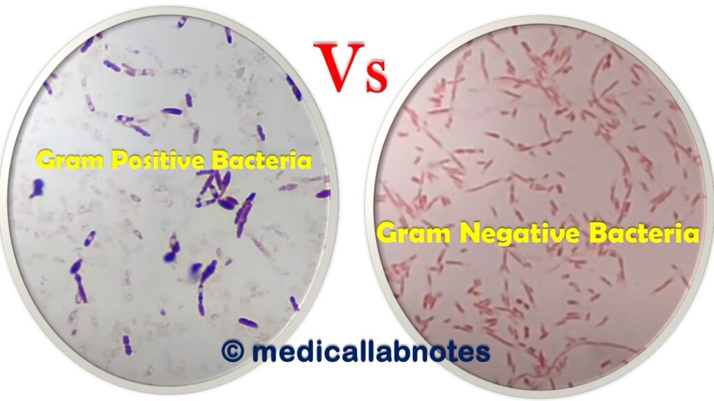
‘Gram-Positive Bacilli (GPB)‘ is also called Gram-Positive Rods (GPR) bacteria which retain crystal violet dye and stain blue or purple on Gram’s staining. The most common medically important bacteria of GPR are Mycobacterium tuberculosis, Mycobacterium leprae, Listeria monocytogenes, Nocardia asteroides, Actinomyces israelii, Bacillus anthracis, Bacillus cereus, Bifidobacterium species, Corynebacterium diphtheriae, and Clostridium species.
‘Gram-Positive Cocci (GPC)‘ bacteria retain crystal violet dye and stain blue or purple on Gram’s staining. The most common medically important bacteria of GPC are Staphylococcus aureus, Streptococcus pneumoniae, Enterococcus species, Streptococcus pyogenes, Streptococcus agalactiae, and Staphylococcus saprophyticus.
‘Gram-Positive Negative (GNB)‘ is also called Gram-Negative Rods (GNR) bacteria which take safranin after decolorization and stain pink or red on Gram’s staining. Most GNR bacteria are medically important and among them, a few are Escherichia coli, Klebsiella pneumoniae, Salmonella enterica serotype Typhi, Shigella species, Proteus species, Citrobacter, Pseudomonas aeruginosa, and Acinetobacter species.
Medically relevant gram-negative cocci bacteria are Neisseria gonorrhoeae (causative agent of sexually transmitted disease), Neisseria meningitidis (causes meningitis), Moraxella catarrhalis (responsible for respiratory symptoms), and Veillonella parvula (anaerobic gram-negative coccus cause osteomyelitis).
Differences between Gram-Positive and Gram-Negative Bacteria
The differences between Gram-Positive and Gram-Negative Bacteria are summarized as follows-
| S. No. | Property | Gram-Positive Bacteria | Gram-Negative Bacteria |
| 1 | Gram Reaction | Take crystal violet (primary stain) and stain violet or purple on staining | Retain safranin (counterstain) after decolorization and stain pink or red on staining |
| 2 | Cell wall thickness | 20-80 nm | 8-10 nm |
| 3 | Peptidoglycan Layer | Multilayered (Thick) | single-layered (Thin) |
| 4 | Rigidity and Elasticity | Rigid and less elastic | Less rigid and more elastic |
| 5 | Outer Membrane | Absent | Present |
| 6 | Variety of amino acid in cell wall | Few | Several |
| 7 | Aromatic and Sulfur-containing amino acid in cell wall | Absent | Present |
| 8 | Periplasmic Space | Absent | Present |
| 9 | Teichoic Acids | Mostly present | Absent |
| 10 | Porins | Absent | Present |
| 11 | Lipopolysaccharide (LPS) Content | Almost None | High |
| 12 | Lipid and Lipoprotein Content | Low (acid-fast bacteria have lipids linked to peptidoglycan) | High (due to presence of outer membrane) |
| 13 | The ratio of RNA and DNA | 8:1 | Nearly 1 |
| 14 | Mesosomes | Fully Prominent | Less Prominent |
| 15 | Flagellar Structure | 2 rings in basal body | 4 rings in basal body |
| 16 | Magnetosomes | Usually absent | Sometimes present |
| 17 | Morphology | Normally cocci or spore-forming rods (except- Lactobacillus and Corynebacterium) | Usually non-spore-forming rods (Except-Neisseria) |
| 18 | Endospore formation | Some produce endospores during unfavorable conditions like Bacillus, Clostridium, Sacchropolyspora, Micromonospora, and Streptomyces. | Normally not producing endospores |
| 19 | Toxin Produced | Exotoxin | Endotoxin or Exotoxin |
| 20 | Pathogens | Only a few are pathogens. | Most of them are pathogens. |
| 21 | Nutritional Requirements | Relatively Complex | Relatively Simple |
| 22 | Resistance to Physical Disruption | High | Low |
| 23 | Cell Wall Disruption by Lysozyme | High | Low ( since it requires pretreatment to destabilize outer membrane) |
| 24 | Susceptibility to Penicillin and Sulfonamide | Sulfonamide High | Low |
| 25 | Susceptibility to Streptomycin, Chloramphenicol, and Tetracycline | Low | High |
| 26 | Inhibition by Basic Dyes | High | Low |
| 27 | Susceptibility to Anionic Detergents | High | Low |
| 28 | Resistance to Sodium Azide | High | Low |
| 29 | Resistance to Drying | High | Low |
| 30 | Rendering | They can render Gram-negative by increasing acidity | They can render Gram-positive by increasing alkalinity |
| 31 | Common Examples | Staphylococcus aureus Streptococcus pnrumoniae Streptococcus pyogenes Streptococcus agalactiae Enterococcus species Bacillus Clostridium Listeria monocytogenes Corynebacterium diphtheriae | Escherichia coli Salmonella Typhi S. Paratyphi, Klebsiella pnumoniae, Proteus vulgaris, Helicobacter pylori Pseudomonas aeruginosa, Acinetobacter spp. |
| 32 | Common Infections | Pneumococcal infections Staphylococcal aureus infections Streptococcal infections Toxic shock syndrome Anthrax Diphtheria Enterococcal infections Erysipelothricosis Listeriosis | E. coli infections, Klebsiella infections, Haemophilus influenzae infections, Cholera, Plague, Typhoid fever, Shigellosis, Brucellosis, Pseudomonas infections, Campylobacter infections |
Gram-Positive and Gram-Negative Bacteria Footages
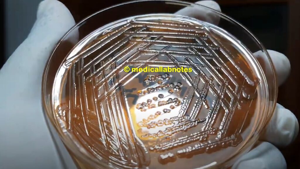
Bacillus species growth on Muller-Hinton Agar

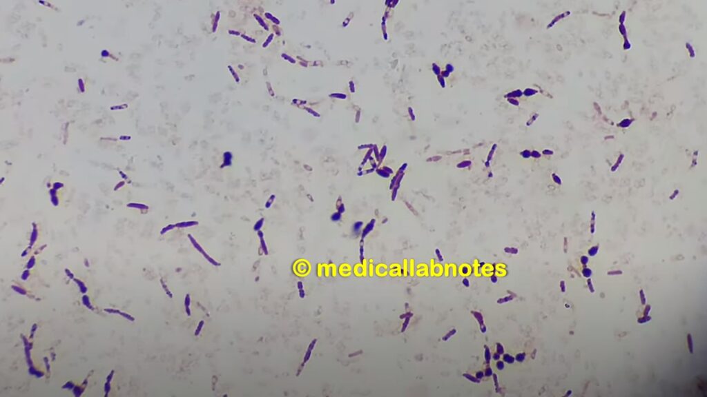
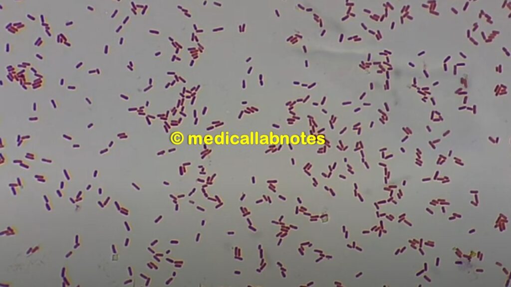
Bacillus species in Gram staining of culture
Mycobacterium tuberculosis in Ziehl-Neelsen staining of Sputum at a high magnification

Mycobacterium leprae in Ziehl-Neelsen staining of Sputum at a high magnification
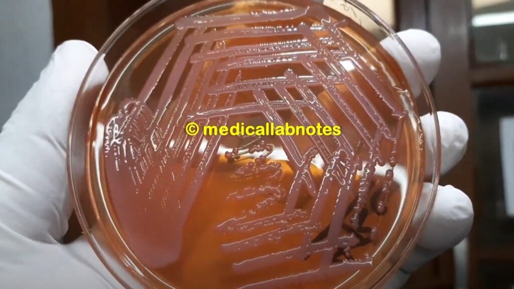
Clostridium growth on blood agar

Clostridium in Gram staining

Listeria monocytogenes colony morphology on blood agar

Listeria monocytogenes in Gram staining of culture

Nocardia in modified Ziehl-Neelsen Staining

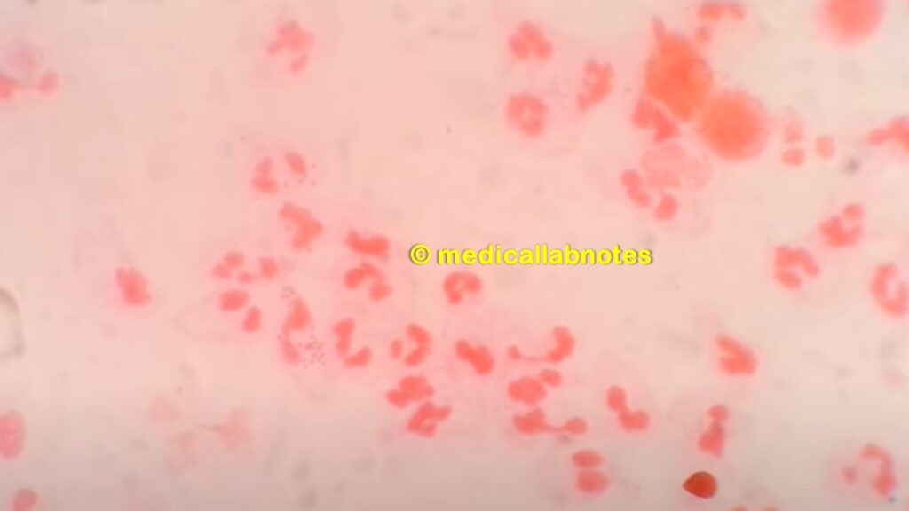
Staphylococcus aureus in Gram staining pus

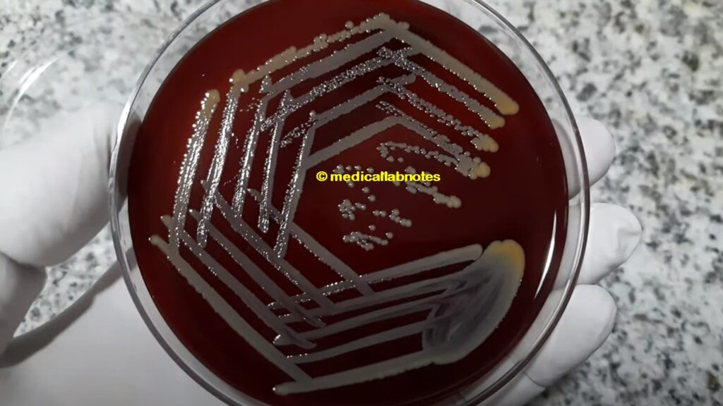
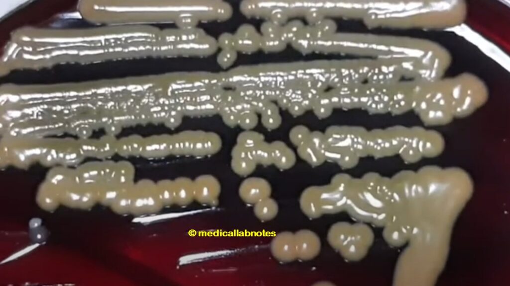
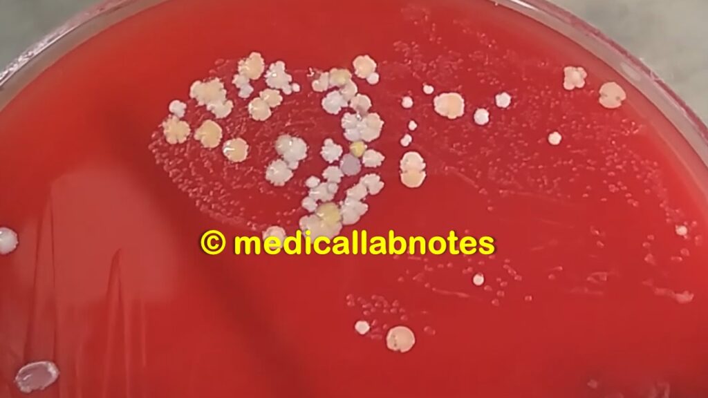
Staphylococcus aureus colony morphology on blood agar
Beta-hemolytic colony of Staphylococcus aureus on blood agar

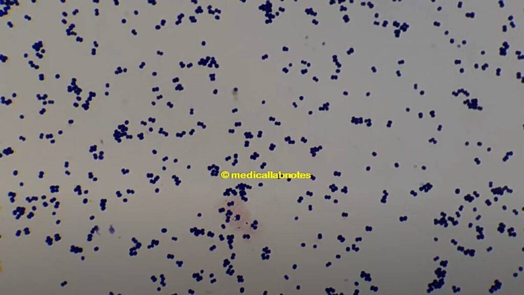
Gram positive cocci in singles, pairs and clusters of Staphylococcus aureus in Gram staining of culture

Micrococcus luteus growth on blood agar
Gram positive cocci in tetrads of Micrococcus
Micrococcus roseus growth on MHA

Micrococcus roseus growth on blood agar

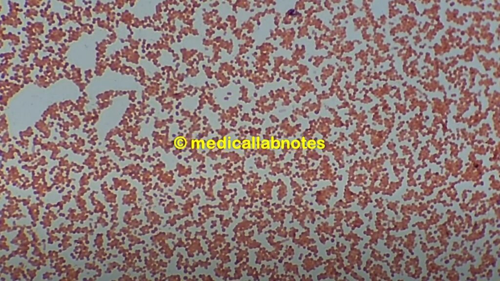
Enterococcus in Gram staining of sputum

Enterococcus growth on blood agar
Enterococcus faecalis in Gram staining of culture microscopy at a high magnification

Enterococcus faecalis colony characteristics on MacConkey agar without bile salt and crystal violet

Enterococcus faecalis colony characteristics on blood agar

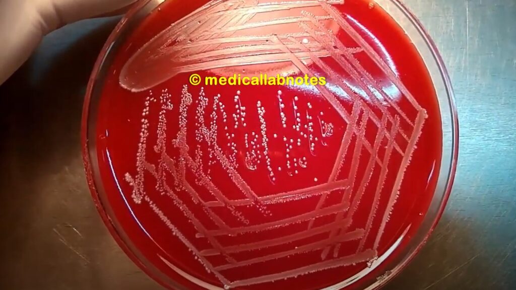
Streptococcus pneumoniae colony morphology on blood agar

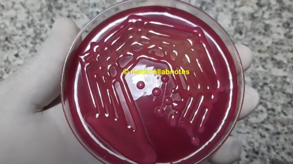
Beta-hemolytic streptococci (Streptococcus pyogenes or Streptococcus agalactiae) colony morphology on blood agar

E. coli colony morphology on MacConkey agar

Escherichia coli in Gram staining of culture

Klebsiella pneumoniae mucoid lactose fermenter colony on MacConkey agar
Klebsiella pneumoniae in Gram staining
Proteus non-lactose fermenter colony on MacConkey medium
Proteus mirabilis in Gram staining of culture

Salmonella Typhi in Gram staining

Pyocyanin and pyoverdin pigments of Pseudomonas aeruginosa

Pseudomonas aeruginosa in Gram staining

Acinetobacter colony morphology on MacConkey agar
Acinetobacter in Gram staining of culture
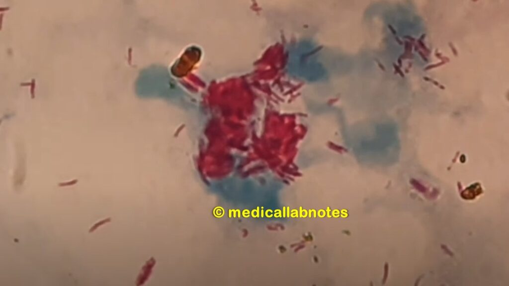
Neisseria gonorrhoeae in Gram staining of urethral discharge showing Gram negative diplococci
Neisseria gonorrhoeae on blood agar of urethral discharge culture
Pure well isolated colony of Neisseria gonorrhoeae on chocolate agar

Neisseria meningitidis growth on blood agar with Micrococcus

Neisseria meningitidis in Gram staining of culture

Further Readings
- https://universe84a.com/gram-stain/
- Jawetz, Melnick and Adelberg’s Medical Microbiology. Editors: Geo. F. Brook, Janet S. Butel & Stephen A. Morse, 21st ed 1998, Publisher Appleton & Lance, Co Stamford Connecticut
- https://connects.catalyst.harvard.edu/Profiles/display/Concept/Gram-Positive%20Endospore-Forming%20Bacteria
- Manual of Clinical Microbiology. Editors: P.R. Murray, E. J. Baron, M. A. Pfaller, F. C. Tenover and R. H. Yolken, 7th ed 2005, Publisher ASM, USA
- https://www.msdmanuals.com/home/infections/bacterial-infections-gram-negative-bacteria/overview-of-gram-negative-bacteria
- Mackie and Mc Cartney Practical Medical Microbiology. Editors: J.G. Colle, A.G. Fraser, B.P. Marmion, A. Simmous, 4th ed, Publisher Churchill Living Stone, New York, Melborne, Sans Franscisco 1996.
- Manual of Clinical Microbiology. Editors: P.R. Murray, E. J. Baron, M. A. Pfaller, F. C. Tenover and R. H. Yolken, 7th ed 2005, Publisher ASM, USA
- Textbook of Diagnostic Microbiology. Editors: Connie R. Mahon, Donald G. Lehman & George Manuselis, 3rd edition2007, Publisher Elsevier.
- Bailey & Scott’s Diagnostic Microbiology. Editors: Bettey A. Forbes, Daniel F. Sahm & Alice S. Weissfeld, 12th ed 2007, Publisher Elsevier.
- Clinical Microbiology Procedure Hand book Chief in editor H.D. Isenberg, Albert Einstein College of Medicine, New York, Publisher ASM (American Society for Microbiology), Washington DC.
- https://www.ncbi.nlm.nih.gov/pmc/articles/PMC180726/
- https://www.asmscience.org/content/education/protocol/protocol.2886
- https://www.sigmaaldrich.com/catalog/product/sigma/ht90a?lang=en
I think this is one of the so much significant info for me. And i am glad studying your article. But should statement on few basic issues, The website style is perfect, the articles is in point of fact great : D. Just right task, cheers
Absolutely composed content, Really enjoyed reading.
Hello very cool site!! Guy .. Beautiful .. Superb .. I’ll bookmark your web site and take the feeds also?KI’m happy to seek out so many useful info here within the post, we need work out extra techniques on this regard, thank you for sharing. . . . . .
Great tremendous things here. I am very glad to peer your article. Thank you so much and i’m looking ahead to touch you. Will you kindly drop me a e-mail?
Howdy very cool web site!! Guy .. Beautiful .. Wonderful .. I’ll bookmark your web site and take the feeds additionally…I am satisfied to find so many useful information here in the publish, we’d like work out extra strategies on this regard, thank you for sharing. . . . . .
very good put up, i actually love this web site, keep on it
What’s Happening i’m new to this, I stumbled upon this I’ve found It positively helpful and it has helped me out loads. I hope to contribute & assist other users like its helped me. Great job.
Excellent weblog here! Additionally your web site lots up fast! What host are you the use of? Can I am getting your associate hyperlink for your host? I wish my website loaded up as fast as yours lol
Only wanna admit that this is very beneficial, Thanks for taking your time to write this.
Hi there, I found your blog by way of Google at the same time as searching for a similar subject, your web site came up, it seems to be great. I have bookmarked it in my google bookmarks.
I cling on to listening to the rumor lecture about getting boundless online grant applications so I have been looking around for the most excellent site to get one. Could you advise me please, where could i acquire some?