
Introduction of Escherichia hermanii
Table of Contents
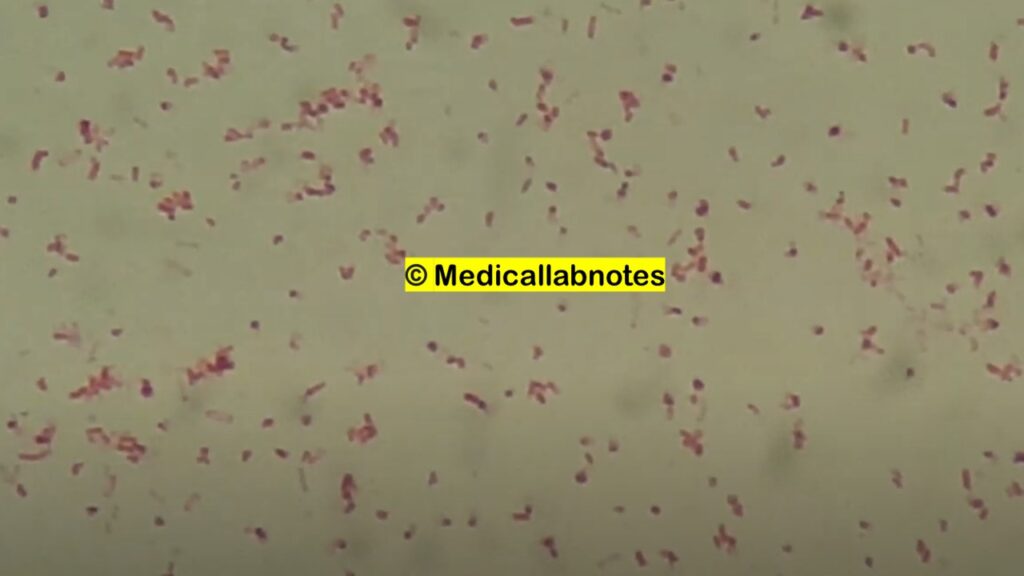
Escherichia hermannii is a gram-negative rod. This bacterium belongs to the family of Enterobacteriaceae. It was first described in 1982. It had been previously known as enteric group 11 of Escherichia coli but was later reclassified as a distinct species in the genus, Escherichia after identifying special genomic features that allowed differentiation from E. coli. In the Clinical Microbiology Laboratory, E. hermannii can be isolated from E. coli having the production of a specific yellow pigment as shown below picture. It is responsible only for rare cases of human infections which are supposed to be mostly a co-infector in polymicrobial infections and it is not considered truly pathogenic. Although, there is evidence of the pathogenicity of this organism, which seems to be able to cause infections even in immunocompetent, and non-predisposed individuals causing bacteremia, urinary tract, and central nervous system infections.

Classiication of Escherichia hermanii
- Domain: Bacteria
- Phylum: Pseudomonadota
- Class: Gammaproteobacteria
- Order: Enterobacterales
- Family: Enterobacteriaceae
- Genus: Escherichia
- Species: E. hermannii
- Binomial name: Escherichia hermannii
Pathogenicity of Escherichia hermanii
- Bacteremia
- Urinary tract infections (UTIs)
- Central nervous system infection
- Gastrointestinal infections
- Peritonitis (abdominal cavity infection)
- Conjunctivitis
- Skin and soft tissue infection (SSTI)
Laboratory Diagnosis of Escherichia hermanii
Biochemical tests of E. hermannii–
- Gram staining -Gram Negative Rods
- Motility -Positive (+)
- Pigment -Positive (+)
- Catalase production -Positive (+)
- Oxidase fermentation -Positive (+)
- Indole-Positive (+)
- Methyl Red-Positive (+)
- Voges-proskauer -Negative (-)
- Citrate -Negative (-)
- Urease -Negative (-)
- H2S Production -Negative (-)
- Oxidase -Negative (-)
- Deoxyribonuclease -Negative (-)
- Lysine decarboxylase -Negative (-)
- Ornithine decarboxylase -Positive (+)
- Arginine dihydrolase -Negative (-)
- Aesculine hydrolysis- d
- Sucrose- d
- Melibiose -Negative (-)
- D-sorbitol -Negative (-)
- Mucate -Positive (+)
- KCN -Positive (+)
- Gelatine -Negative (-)
- Lactose-d
- Phenylalanine deaminase -Negative (-)
- D-mannitol -Positive (+)
- Dulcitol- (-)
- Salicin- d
- Adonitol -Negative (-)
- Raffinose- d
- L-rhamnose -Positive (+)
- Malonate -Negative (-)
- Jordan tartrate- d
- Lipase -Negative (-)
- ONPG -Positive (+)
- D-glucose gas production -Positive (+)
- D-xylose -Positive (+)
- Maltose -Positive (+)
- Cellobiose -Positive (+)
- D-glucose acid production -Positive (+)
- Nitrate reductase -Positive (+)
- Glycerol -Negative (-)
- Acetate utilization – (+)
- L-arabinose -Positive (+)
- Myo-inositol -Negative (-)
- D-Mannose -Positive (+)
- Trehalose -Positive (+)
- Alpha Methyl glucoside -Negative (-)
Keynotes
Positive (+): 90-100% Positive
Negative (-): 0-10% positive
(+): 76-89% positive
(-): 11-25% positive
d: 26-75% positive
v: strain instability

Treatment
Cephalosporins and aminoglycosides are the most common antibiotics used for the treatment of Escherichia hermannii-causing infections.
Keynotes on Escherichia hermanii
- There are also available case reports regarding bloodstream infection by Escherichia hermannii in a neonate and E. hermannii as the sole cause of osteomyelitis in a patient with an open tibial shaft fracture.
- A case report suggests that E. hermanni viscous materials contained mannose-rich exopolysaccharides contributing to its pathogenicity.
E. hermanii in BD™ Tryptic Soy Broth (TSB) wet mount microscopy
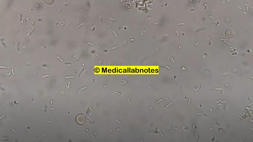
Escherichia hermannii in Wet Mount Microscopy
Bibliography
- https://www.ncbi.nlm.nih.gov/pmc/articles/PMC6473853/
- bioinfo.bisr.res.in/cgi-bin/project/docter/get_details.cgi
- https://en.wikipedia.org/wiki/Escherichia_hermannii
- https://www.jcdr.net/article_fulltext.asp?id=8304
- https://casereports.bmj.com/content/12/11/e231206
- https://academic.oup.com/femspd/article/59/3/456/497186
- https://journals.asm.org/doi/10.1128/JCM.01119-08
I believe people who wrote this needs true loving because
it’s a blessing. So let me give back and speak out on change your life and if you want to seriously get
to hear I will share info about how to learn SNS marketing Don’t forget..
I am always here for yall. Bless yall!
Hello, you used to write wonderful, but the last several posts have been kinda boring?K I miss your tremendous writings. Past few posts are just a little out of track! come on!