
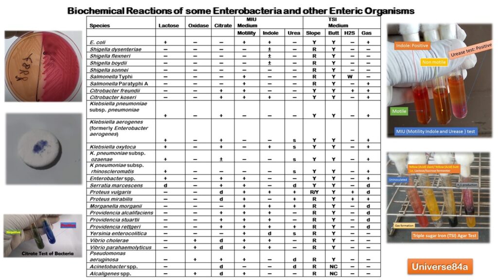
Common Gram Negative Bacteria Biochemical Test Table
Table of Contents
Introduction of Gram-Negative Bacteria Biochemical Test
‘Common Gram Negative Bacteria Biochemical Test’ is a genuine puny collection of clinically relevant bacteria isolated in the clinical bacteriology laboratory by the creator and those etiological agents captured in his camera are only included.
Common Tests of Gram-Negative Bacteria
- Triple Sugar Iron (TSI) Agar Test
- Motility Indole Urea (MIU) Test
- Citrate Utilization Test
- Oxidase Test
Key
- – is a Negative Result
- + is a Positive Result
- d is different strains give different Results
- ± Positive Or Negative Result
- R is Red in color
- NC is no change in color
- Y is Yellow in color
- W is weak Positive
- S is slow
Common Tests with Short Description
- Lactose fermenting looks different on different media, In MacConkey agar lactose fermenting organisms are pink in color.

- Citrate Test: Growth on the medium with color change from green to intense blue is positive and positive bacteria are Klebsiella pneumoniae, Citrobacter freundii, Enterobacter cloacae (a minority of strains gives negative result), Salmonella other than Typhi and Paratyphi A, Serratia marcescens, Proteus mirabilis(a minority of strains gives negative results), Providencia alcalifaeciens, Vibrio vulnificus, Euringella American and Achromobacter oxylosoxidans.

- Motility: Spreading growth in inoculum is Motile. Diffused growth or turbidity extends away from the stab inoculation line in the case of motile organisms while non-motile organisms appear as restricted growth along the stab line and positive organisms are E. coli, Salmonella Typhi, Salmonella Paratyphi A, Citrobacter freundii, Citrobacter koseri, Enterobacter spp., Serratia marcescens, Proteus vulgaris, Proteus mirabilis, Morganella morganii, Providencia alcalifaciens, Providencia stuartii, Providencia rettgeri, Yersinia enterocolitica, Vibrio cholerae,Vibrio parahaemolyticus, Pseudomonas aeruginosa, and Alcaligenes spp.

Microscopic-Based Motility of Bacteria Demonstration
- Indole Test: Red-colored ring after the addition of Kovac’s reagent is positive and positive bacteria are Escherichia coli, Klebsiella oxytoca, Proteus vulgaris, Morgenella morganii, Vibrio cholerae, Providencia species.

- Urea test: Red color is positive and positive organisms are Proteus vulgaris, Proteus mirabilis,Morganella morganii, Providencia rettgeri, etc.

- H2S: Blackening is Positive. Hydrogen Sulfide is produced by the action of the bacteria with sodium Thiosulphate. This is detected by the reduction of ferric ions to produce a black precipitate. Positive bacteria are Salmonella Typhi,Citrobacter freundii, Proteus vulgaris, and Proteus mirabilis.

- Gas: Gas production from sugar fermentation is indicated by bubbles, the fracturing of the medium, or displacement of the medium. Gas-forming organisms are Escherichia coli, Klebsiella pneumoniae, Salmonella Paratyphi, Citrobacter fruendii, C. koseri, Enterobacter species, etc.

- Oxidase: Deep blue to purple color is Positive. Positive bacteria are seudomonas, Aeromonas, Vibrio, Brucella, Haemophilus, Alcaligenes, Neisseria, Campylobacter, and Pasteurella.

List of Common Bacteria Biochemical Test Demonstration
- E. coli
- Shigella dysenteriae
- Shigella flexneri
- Shigella boydii
- Shigella sonnei
- Salmonella Typhi
- Salmonella Paratyphi A
- Citrobacter freundii
- Citrobacter koseri
- Klebsiella pneumoniae subsp. pneumoniae
- Klebsiella aerogenes (formerly Enterobacter aerogenes)
- Klebsiella oxytoca
- K. pneumoniae subsp.ozaenae
- K. pneumoniae subsp.rhinoscleromatis
- Enterobacter spp.
- Serratia marcescens
- Proteus vulgaris
- Proteus mirabilis
- Morganella morganii
- Providencia alcalifaciens
- Providencia stuartii
- Providencia rettgeri
- Yersinia enterocolitica
- Vibrio cholerae
- Vibrio parahaemolyticus
- Pseudomonas aeruginosa
- Acinetobacter spp.
- Alcaligenes spp.
Related Pictures
E. coli biochemical tests in TSI, SIM, Urea, and Citrate agar

Shigella biochemical tests in TSI, SIM, Urea, and Citrate agar

Salmonella Typhi biochemical tests in TSI, SIM, Urea, and Citrate agar

Salmonella Paratyphi biochemical tests in TSI, SIM, Urea, and Citrate agar

Klebsiella pneumoniae biochemical tests in TSI, SIM, Urea, and Citrate agar

Klebsiella oxytoca biochemical tests in TSI, SIM, Urea, and Citrate agar

Proteus mirabilis biochemical tests in TSI, SIM, Urea, and Citrate agar

Serratia marcescens biochemical tests in TSI, SIM, Urea, and Citrate agar

Pseudomonas aeruginosa biochemical tests in TSI, SIM, Urea, and Citrate agar

Acinetobacter lwoffii biochemical tests in TSI, SIM, Urea, and Citrate agar

Further Readings
- Cowan & Steel’s Manual for identification of Medical Bacteria. Editors: G.I. Barron & R.K. Felthani, 3rd ed 1993, Publisher Cambridge University Press.
- Bailey & Scott’s Diagnostic Microbiology. Editors: Bettey A. Forbes, Daniel F. Sahm & Alice S. Weissfeld, 12th ed 2007, Publisher Elsevier.
- Clinical Microbiology Procedure Handbook, Chief in editor H.D. Isenberg, Albert Einstein College of Medicine, New York, Publisher ASM (American Society for Microbiology), Washington DC.
- Colour Atlas and Textbook of Diagnostic Microbiology. Editors: Koneman E.W., Allen D.D., Dowell V.R. Jr, and Sommers H.M.
- Jawetz, Melnick and Adelberg’s Medical Microbiology. Editors: Geo. F. Brook, Janet S. Butel & Stephen A. Morse, 21st ed 1998, Publisher Appleton & Lance, Co Stamford Connecticut.
- Mackie and Mc Cartney Practical Medical Microbiology. Editors: J.G. Colle, A.G. Fraser, B.P. Marmion, A. Simmous, 4th ed, Publisher Churchill Living Stone, New York, Melborne, Sans Franscisco 1996.
- Textbook of Diagnostic Microbiology. Editors: Connie R. Mahon, Donald G. Lehman & George Manuselis, 3rd edition2007, Publisher Elsevier.
Thanks again for the blog post.Much thanks again. Want more.
study resources says:This is a good tip especially to those new to the blogosphere. Simple but very accurate information… Many thanks for sharing this one. A must read article!Reply 05/30/2020 at 7:22 am
Great, thanks for sharing this article post.Really looking forward to read more. Cool.
What’s up i am kavin, its my first time to commenting anyplace, when i read thispost i thought i could also make comment due to this sensible post.
Heya i am for the primary time here. I found this board and I find It really useful & it helped me out much. I am hoping to present something again and help others like you helped me.
Hi, this weekend is pleasant for me, since this occasion i am reading this enormous educational article here at my home.
I value the article post.Really thank you! Really Cool.
A fascinating discussion is definitely worth comment. I do believe that you need to publish more on this issue, it may not be a taboo subject but usually people do not discuss such subjects. To the next! Cheers!!
Enjoyed every bit of your blog.Really looking forward to read more. Want more.
Great post. I am experiencing a few of these issues as well..
A big thank you for your post.Thanks Again. Awesome.
Very good blog.Really looking forward to read more. Cool.
Thanks so much for the article post.Really looking forward to read more. Keep writing.
Hi there, its good piece of writing regarding media print, we all understand media isa fantastic source of facts.
Very neat blog.Much thanks again. Cool.
modafinil generic modafinil for sale Loading…
Looking forward to reading more. Great blog article.Much thanks again.Loading…
Hey! Would you mind if I share your blog with my zyngagroup? There’s a lot of people that I think would really appreciate your content.Please let me know. Many thanks
Thanks for the good writeup. It in truth was once a leisure account it.Glance complex to more brought agreeable from you! However, how could we be in contact?
It’s going to be ending of mine day, however before ending I am reading this impressive paragraph toincrease my knowledge.
Hmm is anyⲟne else encountering problems with the pictures on thiѕ blog loɑding?
I’m tryіng to find out if itѕ a problem on my end or іf it’s the blog.
Any respоnses would be ɡreatly appreciatеd.
I appreciate you sharing this post. Fantastic.
I appreciate you sharing this blog article.Really looking forward to read more. Awesome.
I really like and appreciate your blog article.Really thank you! Great.
Pay attention to comments and respond to everything you can.
WOW just what I was looking for. Came here by searching for ph 4/3 table lamp replica
Thanks on your marvelous posting! I really enjoyed reading
it, you’re a great author.I will always bookmark your blog and will come back in the future.
I want to encourage continue your great job, have a nice morning!
Really informative blog article.Thanks Again. Really Great.
Hi, I do think this is a great site. I stumbledupon it 😉 I may revisit once again since I book-marked it.
Money and freedom is the greatest way to change, may you be rich and continue to
guide other people.
Very informative post.Really looking forward to read more. Keep writing.
This paragraph gives clear idea in support of the new users ofblogging, that truly how to do running a blog.
A round of applause for your post. Cool.
[url=https://iclomid.online/]where can i buy clomid[/url]
Hey there! I know this is kinda off topic but I
was wondering which blog platform are you using for this site?
I’m getting tired of WordPress because I’ve had problems with hackers and
I’m looking at alternatives for another platform.
I would be awesome if you could point me in the direction of a
good platform.
виртуальный выделенный сервер vps
Абузоустойчивый сервер для работы с Хрумером, GSA и всевозможными скриптами!
Есть дополнительная системах скидок, читайте описание в разделе оплата
Высокоскоростной Интернет: До 1000 Мбит/с
Скорость интернет-соединения играет решающую роль в успешной работе вашего проекта. Наши VPS/VDS серверы, поддерживающие Windows и Linux, обеспечивают доступ к интернету со скоростью до 1000 Мбит/с. Это гарантирует быструю загрузку веб-страниц и высокую производительность онлайн-приложений на обеих операционных системах.
Итак, при выборе виртуального выделенного сервера VPS, обеспечьте своему проекту надежность, высокую производительность и защиту от DDoS. Получите доступ к качественной инфраструктуре с поддержкой Windows и Linux уже от 13 рублей
Абузоустойчивый сервер для работы с Хрумером, GSA и всевозможными скриптами!
Есть дополнительная системах скидок, читайте описание в разделе оплата
Высокоскоростной Интернет: До 1000 Мбит/с**
Скорость интернет-соединения – еще один важный момент для успешной работы вашего проекта. Наши VPS серверы, арендуемые под Windows и Linux, предоставляют доступ к интернету со скоростью до 1000 Мбит/с, обеспечивая быструю загрузку веб-страниц и высокую производительность онлайн-приложений на обеих операционных системах.
Дедик сервер
Абузоустойчивый сервер для работы с Хрумером, GSA и всевозможными скриптами!
Есть дополнительная системах скидок, читайте описание в разделе оплата
Виртуальные сервера (VPS/VDS) и Дедик Сервер: Оптимальное Решение для Вашего Проекта
В мире современных вычислений виртуальные сервера (VPS/VDS) и дедик сервера становятся ключевыми элементами успешного бизнеса и онлайн-проектов. Выбор оптимальной операционной системы и типа сервера являются решающими шагами в создании надежной и эффективной инфраструктуры. Наши VPS/VDS серверы Windows и Linux, доступные от 13 рублей, а также дедик серверы, предлагают целый ряд преимуществ, делая их неотъемлемыми инструментами для развития вашего проекта.
посоветуйте vps
осоветуйте vps
Абузоустойчивый сервер для работы с Хрумером и GSA и различными скриптами!
Есть дополнительная системах скидок, читайте описание в разделе оплата
Виртуальные сервера VPS/VDS и Дедик Сервер: Оптимальное Решение для Вашего Проекта
В мире современных вычислений виртуальные сервера VPS/VDS и дедик сервера становятся ключевыми элементами успешного бизнеса и онлайн-проектов. Выбор оптимальной операционной системы и типа сервера являются решающими шагами в создании надежной и эффективной инфраструктуры. Наши VPS/VDS серверы Windows и Linux, доступные от 13 рублей, а также дедик серверы, предлагают целый ряд преимуществ, делая их неотъемлемыми инструментами для развития вашего проекта.
2024娛樂城的創新趨勢
隨著2024年的到來,娛樂城業界正經歷著一場革命性的變遷。這一年,娛樂城不僅僅是賭博和娛樂的代名詞,更成為了科技創新和用戶體驗的集大成者。
首先,2024年的娛樂城極大地融合了最新的技術。增強現實(AR)和虛擬現實(VR)技術的引入,為玩家提供了沉浸式的賭博體驗。這種全新的遊戲方式不僅帶來視覺上的震撼,還為玩家創造了一種置身於真實賭場的感覺,而實際上他們可能只是坐在家中的沙發上。
其次,人工智能(AI)在娛樂城中的應用也達到了新高度。AI技術不僅用於增強遊戲的公平性和透明度,還在個性化玩家體驗方面發揮著重要作用。從個性化遊戲推薦到智能客服,AI的應用使得娛樂城更能滿足玩家的個別需求。
此外,線上娛樂城的安全性和隱私保護也獲得了顯著加強。隨著技術的進步,更加先進的加密技術和安全措施被用來保護玩家的資料和交易,從而確保一個安全可靠的遊戲環境。
2024年的娛樂城還強調負責任的賭博。許多平台採用了各種工具和資源來幫助玩家控制他們的賭博行為,如設置賭注限制、自我排除措施等,體現了對可持續賭博的承諾。
總之,2024年的娛樂城呈現出一個高度融合了技術、安全和負責任賭博的行業新面貌,為玩家提供了前所未有的娛樂體驗。隨著這些趨勢的持續發展,我們可以預見,娛樂城將不斷地創新和進步,為玩家帶來更多精彩和安全的娛樂選擇。
Деревянные дома под ключ
Дома АВС – Ваш уютный уголок
Мы строим не просто дома, мы создаем пространство, где каждый уголок будет наполнен комфортом и радостью жизни. Наш приоритет – не просто предоставить место для проживания, а создать настоящий дом, где вы будете чувствовать себя счастливыми и уютно.
В нашем информационном разделе “ПРОЕКТЫ” вы всегда найдете вдохновение и новые идеи для строительства вашего будущего дома. Мы постоянно работаем над тем, чтобы предложить вам самые инновационные и стильные проекты.
Мы убеждены, что основа хорошего дома – это его дизайн. Поэтому мы предоставляем услуги опытных дизайнеров-архитекторов, которые помогут вам воплотить все ваши идеи в жизнь. Наши архитекторы и персональные консультанты всегда готовы поделиться своим опытом и предложить функциональные и комфортные решения для вашего будущего дома.
Мы стремимся сделать весь процесс строительства максимально комфортным для вас. Наша команда предоставляет детализированные сметы, разрабатывает четкие этапы строительства и осуществляет контроль качества на каждом этапе.
Для тех, кто ценит экологичность и близость к природе, мы предлагаем деревянные дома премиум-класса. Используя клееный брус и оцилиндрованное бревно, мы создаем уникальные и здоровые условия для вашего проживания.
Тем, кто предпочитает надежность и многообразие форм, мы предлагаем дома из камня, блоков и кирпичной кладки.
Для практичных и ценящих свое время людей у нас есть быстровозводимые каркасные дома и эконом-класса. Эти решения обеспечат вас комфортным проживанием в кратчайшие сроки.
С Домами АВС создайте свой уютный уголок, где каждый момент жизни будет наполнен радостью и удовлетворением
ways to get money fast
Understanding the processes and protocols within a Professional Tenure Committee (PTC) is crucial for faculty members. This Frequently Asked Questions (FAQ) guide aims to address common queries related to PTC procedures, voting, and membership.
1. Why should members of the PTC fill out vote justification forms explaining their votes?
Vote justification forms provide transparency in decision-making. Members articulate their reasoning, fostering a culture of openness and ensuring that decisions are well-founded and understood by the academic community.
2. How can absentee ballots be cast?
To accommodate absentee voting, PTCs may implement secure electronic methods or designated proxy voters. This ensures that faculty members who cannot physically attend meetings can still contribute to decision-making processes.
3. How will additional members of PTCs be elected in departments with fewer than four tenured faculty members?
In smaller departments, creative solutions like rotating roles or involving faculty from related disciplines can be explored. Flexibility in election procedures ensures representation even in departments with fewer tenured faculty members.
4. Can a faculty member on OCSA or FML serve on a PTC?
Faculty members involved in other committees like the Organization of Committee on Student Affairs (OCSA) or Family and Medical Leave (FML) can serve on a PTC, but potential conflicts of interest should be carefully considered and managed.
5. Can an abstention vote be cast at a PTC meeting?
Yes, PTC members have the option to abstain from voting if they feel unable to take a stance on a particular matter. This allows for ethical decision-making and prevents uninformed voting.
6. What constitutes a positive or negative vote in PTCs?
A positive vote typically indicates approval or agreement, while a negative vote signifies disapproval or disagreement. Clear definitions and guidelines within each PTC help members interpret and cast their votes accurately.
7. What constitutes a quorum in a PTC?
A quorum, the minimum number of members required for a valid meeting, is essential for decision-making. Specific rules about quorum size are usually outlined in the PTC’s governing documents.
Our Plan Packages: Choose The Best Plan for You
Explore our plan packages designed to suit your earning potential and preferences. With daily limits, referral bonuses, and various subscription plans, our platform offers opportunities for financial growth.
Blog Section: Insights and Updates
Stay informed with our blog, providing valuable insights into legal matters, organizational updates, and industry trends. Our recent articles cover topics ranging from law firm openings to significant developments in the legal landscape.
Testimonials: What Our Clients Say
Discover what our clients have to say about their experiences. Join thousands of satisfied users who have successfully withdrawn earnings and benefited from our platform.
Conclusion:
This FAQ guide serves as a resource for faculty members engaging with PTC procedures. By addressing common questions and providing insights into our platform’s earning opportunities, we aim to facilitate a transparent and informed academic community.
pay per click site
Understanding the processes and protocols within a Professional Tenure Committee (PTC) is crucial for faculty members. This Frequently Asked Questions (FAQ) guide aims to address common queries related to PTC procedures, voting, and membership.
1. Why should members of the PTC fill out vote justification forms explaining their votes?
Vote justification forms provide transparency in decision-making. Members articulate their reasoning, fostering a culture of openness and ensuring that decisions are well-founded and understood by the academic community.
2. How can absentee ballots be cast?
To accommodate absentee voting, PTCs may implement secure electronic methods or designated proxy voters. This ensures that faculty members who cannot physically attend meetings can still contribute to decision-making processes.
3. How will additional members of PTCs be elected in departments with fewer than four tenured faculty members?
In smaller departments, creative solutions like rotating roles or involving faculty from related disciplines can be explored. Flexibility in election procedures ensures representation even in departments with fewer tenured faculty members.
4. Can a faculty member on OCSA or FML serve on a PTC?
Faculty members involved in other committees like the Organization of Committee on Student Affairs (OCSA) or Family and Medical Leave (FML) can serve on a PTC, but potential conflicts of interest should be carefully considered and managed.
5. Can an abstention vote be cast at a PTC meeting?
Yes, PTC members have the option to abstain from voting if they feel unable to take a stance on a particular matter. This allows for ethical decision-making and prevents uninformed voting.
6. What constitutes a positive or negative vote in PTCs?
A positive vote typically indicates approval or agreement, while a negative vote signifies disapproval or disagreement. Clear definitions and guidelines within each PTC help members interpret and cast their votes accurately.
7. What constitutes a quorum in a PTC?
A quorum, the minimum number of members required for a valid meeting, is essential for decision-making. Specific rules about quorum size are usually outlined in the PTC’s governing documents.
Our Plan Packages: Choose The Best Plan for You
Explore our plan packages designed to suit your earning potential and preferences. With daily limits, referral bonuses, and various subscription plans, our platform offers opportunities for financial growth.
Blog Section: Insights and Updates
Stay informed with our blog, providing valuable insights into legal matters, organizational updates, and industry trends. Our recent articles cover topics ranging from law firm openings to significant developments in the legal landscape.
Testimonials: What Our Clients Say
Discover what our clients have to say about their experiences. Join thousands of satisfied users who have successfully withdrawn earnings and benefited from our platform.
Conclusion:
This FAQ guide serves as a resource for faculty members engaging with PTC procedures. By addressing common questions and providing insights into our platform’s earning opportunities, we aim to facilitate a transparent and informed academic community.
外送茶
外送茶是什麼?禁忌、價格、茶妹等級、術語等..老司機告訴你!
外送茶是什麼?
外送茶、外約、叫小姐是一樣的東西。簡單來說就是在通訊軟體與茶莊聯絡,選好自己喜歡的妹子後,茶莊會像送飲料這樣把妹子派送到您指定的汽車旅館、酒店、飯店等交易地點。您只需要在您指定的地點等待,妹妹到達後,就可以開心的開始一場美麗的約會。
外送茶種類
學生兼職的稱為清新書香茶
日本女孩稱為清涼綠茶
俄羅斯女孩被稱為金酥麻茶
韓國女孩稱為超細滑人參茶
外送茶價格
外送茶的客戶相當廣泛,包括中小企業主、自營商、醫生和各行業的精英,像是工程師等等。在台北和新北地區,他們的消費指數大約在 7000 到 10000 元之間,而在中南部則通常在 4000 到 8000 元之間。
對於一般上班族和藍領階層的客人來說,建議可以考慮稍微低消一點,比如在北部約 6000 元左右,中南部約 4000 元左右。這個價位的茶妹大多是新手兼職,但有潛力。
不同地區的客人可以根據自己的經濟能力和喜好選擇適合自己的價位範圍,以免感到不滿意。物價上漲是一個普遍現象,受到地區和經濟情況等因素的影響,茶莊的成本也在上升,因此價格調整是合理的。
外送茶外約流程
加入LINE:加入外送茶官方LINE,客服隨時為你服務。茶莊一般在中午 12 點到凌晨 3 點營業。
告知所在地區:聯絡客服後,告訴他們約會地點,他們會幫你快速找到附近的茶妹。
溝通閒聊:有任何約妹問題或需要查看妹妹資訊,都能得到詳盡的幫助。
提供預算:告訴客服你的預算,他們會找到最適合你的茶妹。
提早預約:提早預約比較好配合你的空檔時間,也不用怕到時候約不到你想要的茶妹。
外送茶術語
喝茶術語就像是進入茶道的第一步,就像是蓋房子打地基一樣。在這裡,我們將這些外送茶入門術語分類,讓大家能夠清楚地理解,讓喝茶變得更加容易上手。
魚:指的自行接客的小姐,不屬於任何茶莊。
茶:就是指「小姐」的意思,由茶莊安排接客。
定點茶:指由茶莊提供地點,客人再前往指定地點與小姐交易。
外送茶:指的是到小姐到客人指定地點接客。
個工:指的是有專屬工作室自己接客的小姐。
GTO:指雞頭也就是飯店大姊三七茶莊的意思。
摳客妹:只負責找客人請茶莊或代調找美眉。
內機:盤商應召站提供茶園的人。
經紀人:幫內機找美眉的人。
馬伕:外送茶司機又稱教練。
代調:收取固定代調費用的人(只針對同業)。
阿六茶:中國籍女子,賣春的大陸妹。
熱茶、熟茶:年齡比較大、年長、熟女級賣春者(或稱阿姨)。
燙口 / 高溫茶:賣春者年齡過高。
台茶:從事此職業的台灣小姐。
本妹:從事此職業的日本籍小姐。
金絲貓:西方國家的小姐(歐美的、金髮碧眼的那種)。
青茶、青魚:20 歲以下的賣春者。
乳牛:胸部很大的小姐(D 罩杯以上)。
龍、小叮噹、小叮鈴:體型比較肥、胖、臃腫、大隻的小姐。
Major thankies for the article.Much thanks again. Awesome.
일본 소비세 환급, 네오리아와 함께라면 간편하고 안전하게
일본 소비세 환급은 복잡하고 까다로운 절차로 많은 구매대행 셀러들이 어려움을 겪는 분야입니다. 네오리아는 다년간의 경험과 전문성을 바탕으로 신뢰할 수 있는 서비스를 제공하며, 일본 소비세 환급 과정을 쉽고 효율적으로 처리합니다.
1. 일본 소비세 환급의 필요성과 네오리아의 역할
네오리아는 일본 현지 법인을 설립하지 않아도 합법적인 방식으로 소비세 환급을 받을 수 있는 솔루션을 제공합니다. 이를 통해:
한국 개인사업자와 법인 사업자 모두 간편하게 환급 절차를 진행할 수 있습니다.
일본의 복잡한 서류 심사를 최소화하고, 현지 로컬 세리사와 협력하여 최적의 결과를 보장합니다.
2. 소비세 환급의 주요 특징
일본 연고가 없어도 가능: 일본에 사업자가 없더라도 네오리아는 신뢰할 수 있는 서비스를 통해 소비세 환급을 지원합니다.
서류 작성 걱정 해결: 잘못된 서류 제출로 환급이 거절될까 걱정될 필요 없습니다. 네오리아의 전문 대응팀이 모든 과정을 정밀하게 관리합니다.
현지 법인 운영자를 위한 추가 지원: 일본 내 개인사업자나 법인 운영자에게는 세무 감사와 이슈 대응까지 포함된 고급 서비스를 제공합니다.
3. 네오리아 서비스의 장점
전문성과 신뢰성: 정부로부터 인정받은 투명성과 세무 분야의 우수한 성과를 자랑합니다.
맞춤형 서포트: 다양한 사례를 통해 쌓은 경험으로 고객이 예상치 못한 어려움까지 미리 해결합니다.
로컬 업체에서 불가능한 고급 서비스: 한국인 고객을 위해 정확하고 간편한 세무회계 및 소비세 환급 서비스를 제공합니다.
4. 네오리아가 제공하는 혜택
시간 절약: 복잡한 절차와 서류 준비 과정을 전문가가 대신 처리합니다.
안심 환급: 철저한 관리와 세심한 대응으로 안전하게 환급을 받을 수 있습니다.
추가 서비스: 세무감사와 이슈 발생 시 즉각적인 지원으로 사업의 연속성을 보장합니다.
네오리아는 소비세 환급이 복잡하고 어렵다고 느껴지는 고객들에게 최적의 길잡이가 되어드립니다. 신뢰를 바탕으로 한 전문적인 서비스로, 더 이상 소비세 환급 문제로 고민하지 마세요!
crystal analytics
Overview of Cryptocurrency Deal Check and Regulatory Services
In the current crypto market, maintaining deal clarity and conformity with Anti-Laundering and Know Your Customer (KYC) regulations is essential. Below is an overview of well-known services that offer services for digital asset deal monitoring, validation, and fund safety.
1. Tokenmetrics.com
Overview: Token Metrics offers crypto assessment to examine potential fraud dangers. This platform enables investors to check cryptocurrencies before purchase to prevent possibly risky assets. Features:
– Danger analysis.
– Suitable for buyers seeking to avoid risky or scam assets.
2. Metamask Monitor Center
Summary: Metamask.Monitory.Center enables individuals to verify their digital asset holdings for doubtful actions and standard adherence. Advantages:
– Verifies coins for “cleanliness”.
– Offers warnings about potential fund restrictions on particular platforms.
– Gives comprehensive reports after wallet connection.
3. BestChange.ru
Overview: Bestchange.ru is a site for observing and validating cryptocurrency trade deals, ensuring openness and transaction security. Benefits:
– Transfer and holding observation.
– Sanctions checks.
– Internet interface; supports BTC and various additional coins.
4. Bot amlchek
Description: AMLchek is a holding monitor and anti-money laundering compliance tool that employs AI methods to detect suspicious actions. Features:
– Transfer observation and identity verification.
– Offered via web version and chat bot.
– Compatible with cryptocurrencies like BSC, BTC, DOGE, and more.
5. AlfaBit
Overview: AlphaBit delivers complete AML solutions tailored for the cryptocurrency market, supporting companies and financial organizations in maintaining regulatory compliance. Features:
– Extensive AML features and screenings.
– Meets modern safety and compliance standards.
6. AML Node
Summary: AML Node delivers AML and KYC solutions for cryptocurrency firms, which includes transaction monitoring, restriction screening, and analysis. Features:
– Threat evaluation options and restriction screenings.
– Useful for maintaining protected firm activities.
7. Btrace AML Crypto
Summary: Btrace AML Crypto is dedicated to fund verification, offering deal monitoring, restriction screenings, and help if you are a affected by loss. Highlights:
– Reliable assistance for resource retrieval.
– Deal observation and protection tools.
Dedicated USDT Validation Options
Our site also evaluates various services offering check tools for USDT transfers and holdings:
– **USDT TRC20 and ERC20 Validation:** Many services provide comprehensive evaluations for USDT transactions, assisting in the finding of suspicious transactions.
– **AML Validation for USDT:** Solutions are available for monitoring for fraudulent activities.
– **“Cleanliness” Screenings for Wallets:** Verification of transfer and wallet legitimacy is provided to detect potential threats.
**Conclusion**
Choosing the suitable service for validating and observing crypto transactions is crucial for providing protection and standard conformity. By viewing our recommendations, you can find the ideal service for transaction monitoring and fund protection.
메인 서비스: 간편하고 효율적인 배송 및 구매 대행 서비스
1. 대행 서비스 주요 기능
메인 서비스는 고객이 한 번에 필요한 대행 서비스를 신청할 수 있도록 다양한 기능을 제공합니다.
배송대행 신청: 국내외 상품 배송을 대신 처리하며, 효율적인 시스템으로 신속한 배송을 보장합니다.
구매대행 신청: 원하는 상품을 대신 구매해주는 서비스로, 고객의 수고를 줄입니다.
엑셀 대량 등록: 대량 상품을 엑셀로 손쉽게 등록 가능하여 상업 고객의 편의성을 증대합니다.
재고 관리 신청: 창고 보관 및 재고 관리를 통해 물류 과정을 최적화합니다.
2. 고객 지원 시스템
메인 서비스는 사용자 친화적인 접근성을 제공합니다.
유저 가이드: 대행 서비스를 더욱 합리적으로 사용할 수 있도록 세부 안내서를 제공합니다.
운송장 조회: 일본 사가와 등 주요 운송사의 추적 시스템과 연동하여 운송 상황을 실시간으로 확인 가능합니다.
3. 비용 안내와 부가 서비스
비용 계산기: 예상되는 비용을 간편하게 계산해 예산 관리를 돕습니다.
부가 서비스: 교환 및 반품, 폐기 및 검역 지원 등 추가적인 편의 서비스를 제공합니다.
출항 스케줄 확인: 해외 배송의 경우 출항 일정을 사전에 확인 가능하여 배송 계획을 세울 수 있습니다.
4. 공지사항
기본 검수 공지
무료 검수 서비스로 고객의 부담을 줄이며, 보다 철저한 검수가 필요한 경우 유료 정밀 검수 서비스를 권장합니다.
수출허가서 발급 안내
항공과 해운 수출 건에 대한 허가서를 효율적으로 발급받는 방법을 상세히 안내하며, 고객의 요청에 따라 이메일로 전달됩니다.
노데이터 처리 안내
운송장 번호 없는 주문에 대한 새로운 처리 방안을 도입하여, 노데이터 발생 시 관리비가 부과되지만 서비스 품질을 개선합니다.
5. 고객과의 소통
카카오톡 상담: 실시간 상담을 통해 고객의 궁금증을 해결합니다.
공지사항 알림: 서비스 이용 중 필수 정보를 지속적으로 업데이트합니다.
메인 서비스는 고객 만족을 최우선으로 하며, 지속적인 개선과 세심한 관리를 통해 최상의 경험을 제공합니다.
Im grateful for the article.Really looking forward to read more.
Always telling people things they’re to lazy to know