
Phenylalanine Deaminase (PDA) Test
Table of Contents
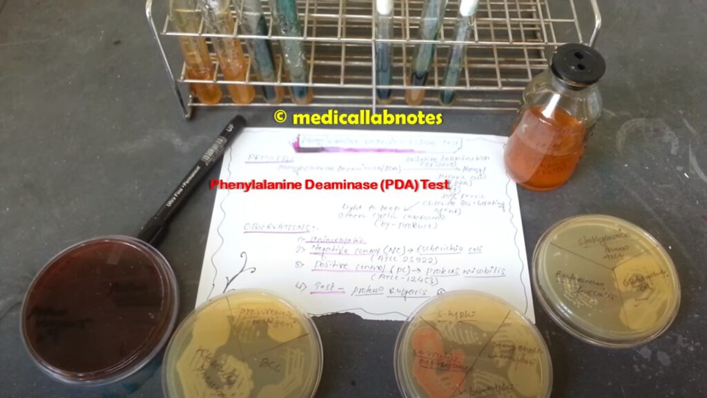
PDA stands for phenylalanine deaminase and PDA tests positive and negative are as shown below footage. It is used in the differentiation of gram-negative enteric bacilli based on the ability of the microorganisms to produce phenyl pyruvic acid by oxidative deamination. The deamination of phenylalanine by oxidative enzymes results in the formation of phenyl pyruvic acid (PPA). After incubation, an aqueous solution of ferric chloride is added. If phenyl pyruvic acid is present, a light to deep green color is produced. Only genera Proteus, Providencia, and Morganella possess enzymes capable of deaminating phenylalanine of the Enterobacteriaceae family.
In 1950, Hendriksen demonstrated that Proteus species were able to convert the amino acid phenylalanine to phenyl pyruvic acid. Later, Buttiaux et al. developed a culture medium for detecting the formation of PPA from phenylalanine by members of the Proteus, Providencia, and Morganella groups. Bynae modified this medium and later Ewing et al. further modified Bynae’s formula by omitting proteose peptone.
Principle of PDA Test
The phenylalanine deaminase (PDA) test is also known as the phenyl pyruvic acid (PPA) test used to differentiate among the urea-positive gram-negative rods based on the ability of the microorganisms to produce PPA by oxidative deamination. Phenylalanine is an amino acid that, upon deamination by oxidase enzymes, results in the formation of phenyl pyruvic acid. The deamination of phenylalanine to PPA is detected by the addition of a ferric chloride solution that acts as a chelating agent with the keto acid by-product to produce a light to deep green cyclic compound. The genus of Enterobacteriaceae like Proteus, Providencia, and Morganella is capable of deaminating phenylalanine and they are also urease-positive and gram-negative rods.
Requirements for Phenylalanine Deaminase (PDA) Test
A. Test organisms:
For the phenylalanine deaminase test take gram-negative rods that grow well on MacConkey agar, are oxidase negative, and are usually urea positive.
B. Media
Phenylalanine agar slants
Composition of the medium
| Ingredients | Gms / Litre |
| DL-Phenylalanine | 2.0 |
| Yeast extract | 3.0 |
| Sodium chloride | 5.0 |
| Disodium phosphate | 1.0 |
| Agar | 12.0 |
| Distilled water | 1000.0 |
| pH | 7.3 |
C. Reagent
10% Ferric chloride, acidified
Composition
| Ingredients | Amount |
| Ferric chloride | 12 gm |
| Concentrated HCl | 2.5 ml |
| Distilled water | 100 ml |
- Dissolve 12 g of ferric chloride in 97.5 ml of water.
- Slowly add 2.5 ml of concentrated HCl to a fume hood.
- Store in a brown bottle at 4°C.
D. Other accessories
- Sterile wooden sticks or inoculating loops
- Incubator
Quality control strains
Organisms for positive control (PC) Proteus mirabilis ATCC 12453 and negative control (NC) Escherichia coli ATCC 25922
Procedure of PDA Test
- Before inoculation, allow the medium to equilibrate at room temperature.
- Using a heavy inoculum from an 18 to 24 hours pure culture, streak the slant surface using a fishtail motion.
- Incubate the inoculated slant aerobically at 37°C for 18 to 24 h. If you are using a heavy inoculum, incubate only for 4 to 6 hours.
- After incubation, apply 4 or 5 drops of ferric chloride directly to the slant.
- Gently roll the reagent over the slant to dislodge surface colonies.
Observation
After the application of ferric chloride, observe the development of a green color within 1 to 5 minutes.
Result and Interpretation of the PDA Test
- Phenylalanine deaminase Test positive: phenylalanine deamination reaction is indicated by the development of light to dark green color within 1 to 5 min after applying ferric chloride reagent.
- Test Negative: Is is indicated by the absence of a green color reaction i.e. negative results will take on a yellow color due to the color of the ferric chloride
- Positive Control: Proteus mirabilis turns green after the addition of ferric chloride
- Negative Control: Escherichia coli remains yellow after the addition of ferric chloride
- Note: The test result is positive as shown above image.
Keynotes of PDA Test
- The test can also use to identify other Enterobacteriaceae, Buttiauxella, Rahnella, and Tatumella, which are PDA positive but are urea negative.
- Tryptophan is also an alternative to phenylalanine; tryptophan deamination releases indole-pyruvic acid, which results in a purple-to-black color with the addition of ferric chloride.
- Either tryptophan or phenylalanine can be used to differentiate among the Proteus group of gram-negative rods.
- The rapid test is available using the disk.
- Many manufacturers follow the formulation established by Ewing.
Limitations of PDA Test
The following are the limitations of the phenylalanine deaminase test:
- The green color reaction of a positive test fades rapidly and therefore, it must interpret within 5 minutes after the application of ferric chloride, or false-negative results may occur.
- Slight agitation of the tube containing ferric chloride will produce a faster, more expressed color reaction.
- Tryptophan deaminase is the substitute for PDA with the same interpretation of results.
- It is recommended that biochemical, immunological, molecular, or mass spectrometry testing be performed on colonies from pure culture for complete identification.
Phenylalanine Deaminase (PDA) Test
- Cowan & Steel’s Manual for identification of Medical Bacteria. Editors: G.I. Barron & R.K. Felthani, 3rd ed 1993, Publisher Cambridge University Press.
- Clinical Microbiology Procedure Handbook, Chief in editor H.D. Isenberg, Albert Einstein College of Medicine, New York, Publisher ASM (American Society for Microbiology), Washington DC.
- Bailey & Scott’s Diagnostic Microbiology. Editors: Bettey A. Forbes, Daniel F. Sahm & Alice S. Weissfeld, 12th ed 2007, Publisher Elsevier.
- Colour Atlas and Textbook of Diagnostic Microbiology. Editors: Koneman E.W., Allen D.D., Dowell V.R. Jr, and Sommers H.M.
- Jawetz, Melnick and Adelberg’s Medical Microbiology. Editors: Geo. F. Brook, Janet S. Butel & Stephen A. Morse, 21st ed 1998, Publisher Appleton & Lance, Co Stamford Connecticut.
- https://catalog.hardydiagnostics.com/cp_prod/Content/hugo/PhenylalanineAgar.htm
- Mackie and Mc Cartney Practical Medical Microbiology. Editors: J.G. Colle, A.G. Fraser, B.P. Marmion, A. Simmous, 4th ed, Publisher Churchill Living Stone, New York, Melbourne, Sans Francisco 1996.
- Textbook of Diagnostic Microbiology. Editors: Connie R. Mahon, Donald G. Lehman & George Manuselis, 3rd edition2007, Publisher Elsevier.
At this time it looks like BlogEngine is the best blogging platform available right now. (from what I’ve read) Is that what you’re using on your blog?