
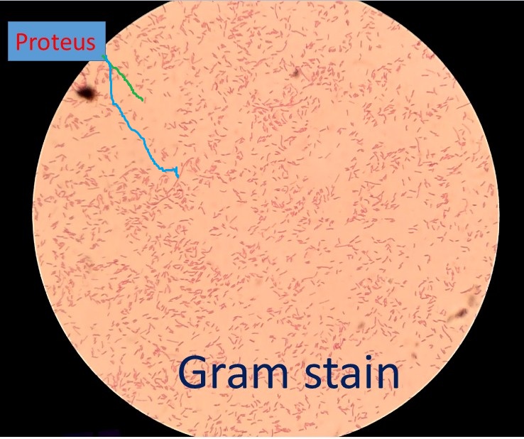
The genus Proteus was discovered in 1885 by Hauser and it is also named after a Greek god. Proteus is a member of the family, Enterobacteriaceae and it is a Gram-negative, oxidase-negative, fimbriated, motile, non-sporing rod-shaped bacterium without capsule and having a size of 0.4–0.8 μm in diameter and 1.0–3.0 μm in length. Proteus vulgaris is naturally found in the natural environment and also in the intestinal tract.

It is a gut bacterium inside our intestines whereas, outside the gut, it can cause serious infections. It is an etiological agent of catheter-associated UTI (CAUTI), sepsis, and septic shock if the infection goes up and causes cystitis and pyelonephritis.

Biochemical Reactions of Proteus vulgaris
Table of Contents
| Basic Features | Properties |
| 1. Gram Staining | Gram-Negative Rods (GNRs) |
| 2. Spore | Non-Sporing |
| 3. Capsule | Negative |
| 4. Motility | Motile |
| 5. Pigment | Negative |
| 6. Growth in potassium cyanide (KCN) medium | Positive |
| 7. Catalase test | Positive |
| 8. Oxidase test | Negative |
| 9. Nitrate reduction test | Positive |
| 10. MR (Methyl Red) test | Positive |
| 11. VP (Voges- Proskauer) assay | Negative |
| 12. OF (Oxidative-Fermentative) test | Fermentative (facultative anaerobes) |
| 13. Gas from Glucose | Positive |
| 14. H2S production | Positive |
| 15. Indole formation | Negative |
| 16. Urease/ urea hydrolysis test | Positive |
| 17. Citrate/citrate utilization | Positive |
| 18. DNase test | Variable |
| 19. Glucose fermentation | Positive |
| 20. Maltose fermentation | Negative |
| 21. Lactose fermentation | Negative |
| 22. Sucrose fermentation | Negative |
| 23. Xylose fermentation | Positive |
| 24. Mannitol fermentation | Negative |
| 25. Acetate Utilization | Negative |
| 26. ONPG (β-galactosidase) | Negative |
| 27. Phenylalanine Deaminase (PDA)/PPA Test | Positive |
| 28. Lipase test | Positive |
| 29. Esculin Hydrolysis test | Negative |
| 30. Lysine Decarboxylase Test | Negative |
| 31. Ornithine Decarboxylase Test | Positive |
| 32. Arginine Dihydrolase Test | Negative |
| 33. Gelatin Hydrolysis | Positive |
| 34. Tryptophan Deaminase | Negative |
| 35. Casein Hydrolysis | Negative |
Keynotes on Proteus
- In Greek mythology, Proteus means sea god.
- Every year about 150 million people are affected by Proteus mirabilis globally.
- It is the bacterium of concern since, in the USA, it accounts for about 3% of all hospital infections and 44% of CAUTI.
- The principal virulence factors associated with infection are flagella, pili, urease, hemolysin, and metal intake.
- Multiple drug-resistant (MDR) strains to carry R plasmids have become very important in nosocomial infections.
- The distinctive characteristics of the genus are PPA, urease, and H2S positive.
- Indole helps to differentiate P. vulgaris (positive) from P. mirabilis ( negative).
- Dienes phenomenon or typing is used successfully to determine the relationship between strains of Proteus species in studies of cross-infection.
- The swarming growth of Proteus contains swimmer and swarmer cells and these cells can be determined using Gram’s staining i.e. swimmer cells- small-near the center of the growth plate while swarmer cells- large-away the center of the growth plate as shown in footages.

- The swarming growth of Proteus is inhibited by the following agents-
- Agar (6%)
- Sodium azide (NaN3) (1:500)
- Chloral hydrate (1:500)
- Boric acid (1:1000)
- Alcohol (5-6%)
Proteus Footages
Swarming growth of Proteus on blood agar

Proteus in Gram Staining
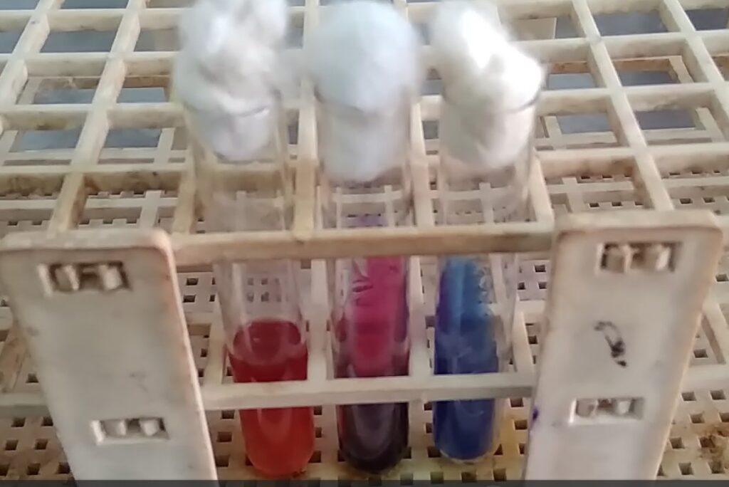
Proteus vulgaris Biochemical Tests-MIU, TSI, and Citrate Utilization Tests
Dienes phenomenon of Proteus vulgaris strains

Proteus mirabilis Biochemical Tests-MIU, TSI, and Citrate Utilization Tests

Further Reading
- https://jb.asm.org/content/195/6/1305
- https://www.karger.com/Article/FullText/514097
- https://www.annualreviews.org/doi/pdf/10.1146/annurev.mi.32.100178.000533
- https://universe84a.com/proteus-general-characteristics
- Williams FD, Schwarzhoff RH. 1978. Nature of the swarming phenomenon in Proteus. Annu. Rev. Microbiol. 32:101–122.
- https://www.sciencedirect.com/topics/medicine-and-dentistry/proteus-mirabilis
- Armbruster CE, Mobley HLT. 2012. Merging mythology and morphology: the multifaceted lifestyle of Proteus mirabilis. Nat. Rev. Microbiol. 10:743–754
- Morgenstein RM, Szostek B, Rather PN. 2010. Regulation of gene expression during swarmer cell differentiation in Proteus mirabilis. FEMS Microbiol. Rev. 34:753–763.
- Rather PN. 2005. Swarmer cell differentiation in Proteus mirabilis. Environ. Microbiol. 7:1065–1073.
- Bailey & Scott’s Diagnostic Microbiology. Editors: Bettey A. Forbes, Daniel F. Sahm & Alice S. Weissfeld, 12th ed 2007, Publisher Elsevier.
- Clinical Microbiology Procedure Handbook, Chief in editor H.D. Isenberg, Albert Einstein College of Medicine, New York, Publisher ASM (American Society for Microbiology), Washington DC.
- Colour Atlas and Textbook of Diagnostic Microbiology. Editors: Koneman E.W., Allen D.D., Dowell V.R. Jr, and Sommers H.M.
- Textbook of Diagnostic Microbiology. Editors: Connie R. Mahon, Donald G. Lehman & George Manuselis, 3rd edition 2007, Publisher Elsevier
- Jawetz, Melnick and Adelberg’s Medical Microbiology. Editors: Geo. F. Brook, Janet S. Butel & Stephen A. Morse, 21st ed 1998, Publisher Appleton & Lance, Co Stamford Connecticut.
- Mackie and Mc Cartney Practical Medical Microbiology. Editors: J.G. Colle, A.G. Fraser, B.P. Marmion, A. Simmous, 4th ed, Publisher Churchill Living Stone, New York, Melborne, Sans Franscisco 1996.
- Manual of Clinical Microbiology. Editors: P.R. Murray, E. J. Baron, M. A. Pfaller, F. C. Tenover and R. H. Yolken, 7th ed 2005, Publisher ASM, USA
I may need your help. I tried many ways but couldn’t solve it, but after reading your article, I think you have a way to help me. I’m looking forward for your reply. Thanks.
Hi, its pleasant piece of writing concerning media print, we all know media is a impressive source of information.
Usually I do not read article on blogs, however I would like to say that
this write-up very forced me to check out and do it!
Your writing taste has been surprised me. Thanks, quite great post.
Hi my loved one! I wish to say that this post is amazing, nice written and come with approximately all significant infos. I would like to see extra posts like this.