
Introduction
Table of Contents
Pulsed-field gel electrophoresis (PFGE) is a powerful laboratory technique used in molecular biology and genetics to separate and analyze large DNA molecules, such as genomic DNA, by their size. PFGE is particularly valuable when studying organisms with complex genomes, such as bacteria, yeast, and higher eukaryotes, because it allows for the resolution of extremely large DNA fragments, up to several megabases in size.
Here is an introduction to the key aspects of PFGE:
- Principle: PFGE employs an alternating electric field, which periodically changes direction, to manipulate the movement of DNA molecules within an agarose gel matrix. The direction and strength of the field are controlled to ensure that DNA fragments of varying sizes migrate through the gel at different rates.
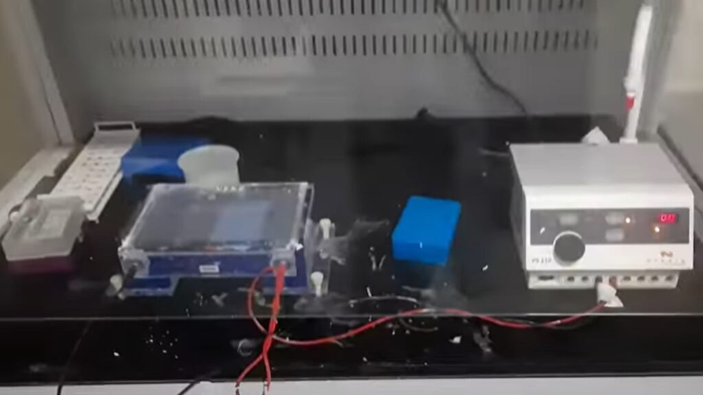
- Equipment: PFGE requires specialized equipment, including a pulse generator, gel electrophoresis chamber, and a cooling system. The gel chamber is typically designed to accommodate large gels and electrodes placed at both ends to create the pulsed electric field.
- Sample Preparation: Before PFGE, the DNA sample of interest is usually embedded within the gel. This is done by mixing the DNA with a molten agarose solution and then pouring it into a mold to solidify. The resulting gel block containing the DNA is then loaded into the electrophoresis chamber.
- Pulsed Electrophoresis: During PFGE, the direction of the electric field alternates at regular intervals, causing the DNA molecules to repeatedly switch directions as they move through the gel. This periodic reversal of the field helps prevent the DNA from becoming trapped or migrating in a single direction. It allows even the largest DNA fragments to navigate the gel matrix effectively.
- Separation: The DNA molecules separate based on their size, with larger fragments moving more slowly through the gel than smaller ones. The electrophoretic mobility of DNA in PFGE is influenced by factors like pulse duration, voltage, and the angle of the field, which can be adjusted to optimize resolution.
- Visualization and Analysis: After electrophoresis, the DNA fragments are typically stained with a DNA-specific dye and visualized using ultraviolet (UV) light. The resulting pattern of DNA bands on the gel provides information about the size distribution of the DNA fragments in the sample.
- Applications:
- Genomic Mapping: PFGE is commonly used for mapping the genomes of various organisms, aiding in the identification and characterization of genes, restriction fragment length polymorphisms (RFLPs), and other genetic elements.
- Epidemiology: It plays a crucial role in epidemiological investigations, helping to track the spread of bacterial pathogens and identify the source of outbreaks.
- Genetic Fingerprinting: PFGE is used in DNA fingerprinting, forensic analysis, and paternity testing.
- Comparative Genomics: Researchers use PFGE to compare the genomes of different organisms or strains, providing insights into genetic variations and evolution.
Principle
The principle of Pulsed-field gel electrophoresis (PFGE) revolves around the use of a pulsing electric field to separate and analyze large DNA molecules based on their size. This technique overcomes the limitations of traditional gel electrophoresis, which is more suitable for smaller DNA fragments. Here’s a detailed explanation of the principle of PFGE:
- Alternating Electric Field: PFGE employs an alternating electric field that periodically changes direction. Unlike conventional gel electrophoresis, where the electric field remains constant, the alternating field in PFGE is a critical aspect of its principle.
- DNA Migration: When DNA molecules are subjected to this alternating electric field, they migrate through a gel matrix. The gel is typically made of agarose or polyacrylamide, and it provides a porous medium for DNA to traverse.
- Directional Changes: The key feature of PFGE is that the direction of the electric field alternates at regular intervals, causing the DNA molecules to periodically switch directions as they move through the gel. This periodic reversal of the field helps prevent the DNA from migrating in a single direction, allowing even the largest DNA fragments to navigate the gel effectively.
- Size-Based Separation: As DNA fragments migrate through the gel, they encounter resistance due to the pores in the matrix. The larger DNA fragments experience greater resistance and move more slowly, while smaller fragments can traverse the gel more quickly. Consequently, DNA fragments are separated in the gel based on their size.
- Pulse Duration and Field Strength: The resolution and separation of DNA fragments in PFGE can be controlled by adjusting parameters such as the pulse duration (the time the field is on) and the field strength (the voltage applied). By modifying these parameters, researchers can optimize the separation of DNA fragments of different sizes.
- Visualization and Analysis: After PFGE is complete, the gel is typically stained with a DNA-specific dye, and the resulting DNA bands are visualized under ultraviolet (UV) light. The pattern of bands on the gel corresponds to the distribution of DNA fragment sizes in the sample. Larger DNA fragments appear closer to the origin (where the DNA was loaded), while smaller fragments migrate farther.
- Quantitative Analysis: PFGE can be used for quantitative analysis by comparing the position and intensity of DNA bands with known standards or markers. This information can be used to estimate the sizes of unknown DNA fragments.
Test Requirements for Pulsed-field gel electrophoresis (PFGE)
Performing Pulsed-field gel electrophoresis (PFGE) requires several specific laboratory and equipment requirements to ensure accurate and successful results. Here are the key test requirements for PFGE:
- Electrophoresis Apparatus: You will need a specialized electrophoresis chamber designed for PFGE. This apparatus should have the capability to generate a pulsed electric field, control pulse duration and voltage, and accommodate large agarose or polyacrylamide gels. It should also include electrodes positioned at both ends to create the pulsed field.
- Cooling System: PFGE generates a significant amount of heat due to the long electrophoresis run times and the high voltage required. Therefore, a cooling system is essential to maintain a stable gel temperature during the procedure. This can include a recirculating water bath or a built-in cooling system in the PFGE apparatus.
- Agarose or Polyacrylamide Gel: You’ll need a gel matrix for electrophoresis. Agarose gels are commonly used for PFGE due to their ability to separate large DNA fragments effectively. The gel concentration can vary depending on the size range of DNA fragments you intend to separate.
- DNA Samples: Prepare your DNA samples of interest. These could be genomic DNA, plasmid DNA, or other types of DNA that you want to separate and analyze. Ensure that your DNA is of high quality and adequately quantified.
- Restriction Endonucleases: To create a molecular weight marker or restriction fragment patterns, you may need restriction enzymes to digest the DNA samples. Select restriction enzymes that will produce suitable fragments for your analysis.
- Loading Buffer: A loading buffer is used to mix with your DNA samples before loading them into the gel. This buffer contains tracking dyes that help visualize the DNA migration during electrophoresis.
- Pulse Generator: A pulse generator or programmable power supply is necessary to create the alternating electric field required for PFGE. This device allows you to set the pulse duration, voltage, and pulse angle.
- Electrodes: High-quality electrodes are needed to apply the pulsed electric field across the gel. Ensure they are properly positioned and in good condition to avoid electrical arcing or other issues during the procedure.
- Power Supply: A stable power supply capable of delivering the required voltage and current is essential for PFGE. This power supply should be compatible with the electrophoresis apparatus.
- UV Transilluminator: After PFGE, you’ll need a UV transilluminator to visualize the DNA bands on the gel. This is typically used in combination with DNA-specific dyes to stain the gel.
- Gel Documentation System: For documentation and analysis of PFGE results, a gel documentation system equipped with a camera and software for image capture and analysis is beneficial.
- Safety Equipment: Ensure you have appropriate safety equipment, such as lab coats, gloves, and safety goggles, as PFGE involves working with electrical equipment and potentially hazardous chemicals.
- Standard DNA Markers: Standard DNA markers with known fragment sizes should be included on the gel to create a molecular weight reference ladder for estimating the sizes of DNA fragments in your samples.
- Quality Control: Regularly calibrate and quality control your equipment, including the electrophoresis apparatus, power supply, and cooling system, to ensure consistent and reliable results.
- Proper Training: Personnel conducting PFGE should be properly trained in the technique to ensure safe and accurate execution of the procedure.
Procedure
Performing Pulsed-field gel electrophoresis (PFGE) requires a detailed procedure to separate and analyze large DNA fragments based on their size. Here is a general step-by-step procedure for PFGE:
Note: The specific details of the procedure may vary depending on the equipment and reagents you are using, so always refer to the manufacturer’s instructions and protocols for your particular setup.
Materials and Reagents:
- Agarose gel (low-melting-point agarose is commonly used)
- DNA samples (genomic DNA or other large DNA fragments)
- Restriction enzymes (if needed for genomic DNA digestion)
- Electrophoresis buffer (usually 0.5x TBE or 0.5x TAE)
- Loading buffer with DNA-specific dye
- DNA molecular weight marker
- PFGE electrophoresis apparatus with cooling system
- Pulse generator or power supply
- UV transilluminator
- Gel documentation system
Procedure:
- Preparation of Gel:
- Prepare an agarose gel with an appropriate concentration based on the size range of DNA fragments you want to separate. Usually, a 0.5% to 2% agarose gel is suitable.
- Cast the gel in a mold that fits your electrophoresis chamber. Ensure there are wells for loading samples.
- Add ethidium bromide or a similar DNA-specific stain to the gel to make DNA bands visible during UV visualization.
- Preparation of DNA Samples:
- If working with genomic DNA, digest it with an appropriate restriction enzyme(s) to create fragments of interest. This step is optional and depends on your research goals.
- Purify the DNA fragments to remove any contaminants or residual enzymes.
- Quantify the DNA concentration accurately.
- Loading Samples:
- Mix each DNA sample with an appropriate volume of loading buffer containing a DNA-specific dye.
- Load the DNA samples into the wells of the agarose gel.
- Setting Up the PFGE Apparatus:
- Place the gel in the PFGE electrophoresis chamber, ensuring it is properly seated.
- Fill the electrophoresis chamber with 0.5x TBE or 0.5x TAE electrophoresis buffer.
- Insert electrodes at both ends of the gel and ensure they are securely in place.
- Applying the Pulsed Electric Field:
- Set the pulse generator or power supply to your desired parameters, including pulse duration, voltage, and angle of the field. These parameters may need to be optimized for your specific experiment.
- Start the electrophoresis run. The alternating electric field will cause the DNA fragments to migrate through the gel.
- Electrophoresis Run:
- Run the PFGE for an appropriate amount of time, which can vary depending on your samples and settings. PFGE runs can take several hours to complete.
- UV Visualization:
- After the run is complete, carefully remove the gel from the chamber.
- Place the gel on a UV transilluminator to visualize the DNA bands. The DNA-specific dye will fluoresce under UV light, allowing you to see the bands.
- Image Documentation:
- Capture an image of the gel using a gel documentation system for further analysis.
- Data Analysis:
- Analyze the gel image to determine the sizes and patterns of the separated DNA fragments. You can compare them to the molecular weight marker for size estimation.
- Cleanup and Disposal:
- Properly dispose of the agarose gel, buffer, and any potentially hazardous materials used in the procedure according to your laboratory’s waste disposal guidelines.
Result-Interpretation of Pulsed-field gel electrophoresis (PFGE)
Interpreting the results of Pulsed-field gel electrophoresis (PFGE) involves analyzing the pattern of DNA bands on the gel to gain insights into the size distribution of the separated DNA fragments. Here’s how to interpret PFGE results:
- Visualization: After completing the PFGE run and staining the gel with a DNA-specific dye, you’ll observe a pattern of DNA bands when you place the gel on a UV transilluminator. These bands represent the separated DNA fragments.
- Molecular Weight Marker: To interpret the size of the DNA fragments, compare the positions of the bands on the gel to a molecular weight marker that was run alongside your samples. The marker typically contains DNA fragments of known sizes. The distances the bands travel in comparison to the marker will provide an estimate of the sizes of your DNA fragments.
- Pattern Analysis:
- Number of Bands: Count the number of distinct bands in your samples. Each band represents a different DNA fragment.
- Intensity of Bands: The intensity of a band indicates the relative amount of DNA in that fragment. More intense bands contain more DNA.
- Mobility of Bands: Larger DNA fragments will migrate more slowly through the gel and appear closer to the origin (where the DNA was loaded), while smaller fragments will migrate farther.
- Fragment Sizes:
- Use the molecular weight marker as a reference to estimate the sizes of your DNA fragments. You can create a standard curve by plotting the known sizes of the marker fragments against the distances they traveled. Then, measure the distances traveled by your sample bands and estimate their sizes from the standard curve.
- Some PFGE software packages can automate this process by analyzing the gel image and providing size estimates for the bands.
- Pattern Comparison:
- If you’re conducting comparative studies, compare the PFGE patterns between different samples or strains. Differences in band patterns can provide valuable information about genetic relatedness or differences in DNA content.
- Similarities in band patterns might indicate a common genetic origin or close genetic relationship, while differences can suggest genetic variation or divergence.
- Quality Control: Assess the overall quality of your PFGE run. Ensure that the bands are well-separated and distinct. Overloaded or underloaded gels, improper buffer conditions, or equipment issues can result in poor-quality PFGE results.
- Documentation: Document your results by capturing images of the gel using a gel documentation system. Include information about the samples, the molecular weight marker used, and any experimental conditions.
- Data Interpretation: Interpret the results in the context of your research goals. For example, in epidemiology, PFGE patterns might be used to trace the source of a bacterial outbreak, while in genomics, they might help in genome mapping or characterizing genetic variation.
- Reporting: Summarize your findings in a clear and concise manner in your research report or publication. Include any relevant statistical analyses if applicable.
Application
Pulsed-field gel electrophoresis (PFGE) is a versatile molecular biology technique that finds applications in various fields of research and diagnostics due to its ability to separate and analyze large DNA fragments based on size. Some of the prominent applications of PFGE include:
- Genomic Mapping:
- Bacterial Genomic Mapping: PFGE is widely used for mapping the genomes of bacteria. It helps in determining the order and relative positions of various genes and genetic elements within a bacterial chromosome. This information is essential for understanding bacterial genetics and genomics.
- Epidemiology:
- Outbreak Investigations: In epidemiology, PFGE is crucial for tracking the source of bacterial outbreaks, such as foodborne illnesses and hospital-acquired infections. By comparing the DNA patterns of bacterial strains, epidemiologists can identify common sources and transmission routes.
- Genetic Fingerprinting:
- Forensic Analysis: PFGE is employed in forensic DNA analysis to create genetic profiles of individuals or samples. It can help in solving criminal cases, identifying victims, and establishing relationships in paternity and maternity testing.
- Comparative Genomics:
- Genomic Variation Studies: Researchers use PFGE to compare the genomes of different organisms or strains. This allows for the identification of genetic variations, such as insertions, deletions, and rearrangements. Comparative genomics is valuable for understanding evolutionary relationships and adaptation.
- Plasmid Analysis:
- Plasmid Typing: PFGE is used to characterize plasmids, which are small, circular DNA molecules often found in bacteria. Plasmid analysis helps in understanding plasmid diversity and tracking the spread of antibiotic resistance genes among bacterial populations.
- Molecular Typing:
- Molecular Epidemiology: PFGE is a key tool in molecular epidemiology, where it helps in identifying strains of pathogens responsible for infectious diseases. This information aids in tracking the spread of diseases and designing effective control measures.
- Environmental Microbiology:
- Water Quality Assessment: PFGE can be used to analyze microbial communities in water sources. It helps in monitoring the presence and distribution of specific microorganisms, including pathogens and indicator organisms, in environmental samples.
- Genome Assembly:
- Genome Sequencing: In genome sequencing projects, PFGE is employed to create high-molecular-weight DNA fragments that serve as the starting material for the sequencing process. These large fragments facilitate genome assembly.
- Agriculture and Food Safety:
- Foodborne Pathogen Characterization: PFGE is used to characterize foodborne pathogens like Salmonella, Escherichia coli (E. coli), and Listeria. This is crucial for food safety and traceability in the food industry.
- Cancer Research:
- Genomic Analysis: PFGE has applications in cancer research, where it can be used to study DNA fragments associated with cancerous cells, such as DNA from tumor samples or fragments generated during chromosomal translocations.
- Diagnostic Laboratories:
- Clinical Diagnostics: PFGE can be employed in clinical settings to characterize pathogens and assess their relatedness in cases of nosocomial infections, disease outbreaks, or when tracking the spread of antibiotic resistance genes.
Keynotes
Here are some keynotes on Pulsed-field gel electrophoresis (PFGE):
- Large DNA Molecule Separation: PFGE is a technique used to separate and analyze large DNA molecules, including genomic DNA, by size. It can resolve DNA fragments ranging from tens of kilobases to several megabases.
- Alternating Electric Field: PFGE uses an alternating electric field that periodically changes direction to manipulate the movement of DNA molecules within an agarose gel matrix. This alternating field allows even the largest DNA fragments to migrate effectively.
- Specialized Equipment: Performing PFGE requires specialized equipment, including a PFGE electrophoresis chamber, pulse generator, and cooling system to maintain gel temperature during long runs.
- Molecular Weight Marker: A molecular weight marker containing DNA fragments of known sizes is typically run alongside samples to estimate the sizes of DNA fragments in the samples.
- Applications:
- PFGE has a wide range of applications, including genomic mapping, epidemiology (outbreak investigations), genetic fingerprinting, comparative genomics, plasmid analysis, and molecular epidemiology.
- It is used in fields such as microbiology, genetics, genomics, forensics, and environmental science.
- Pattern Analysis: PFGE results in a pattern of DNA bands on a gel. The number, position, and intensity of these bands provide information about the size distribution and quantity of DNA fragments in the sample.
- Molecular Epidemiology: In epidemiology, PFGE is crucial for tracking the source and transmission of infectious diseases, making it an essential tool in public health investigations.
- Genome Mapping: PFGE is commonly used for bacterial genome mapping, aiding in the identification and characterization of genes, genomic rearrangements, and other genetic elements.
- Forensic DNA Analysis: PFGE is utilized in forensic science to create genetic profiles of individuals and analyze DNA evidence in criminal investigations.
- Environmental Monitoring: In environmental microbiology, PFGE can help assess water quality by detecting and characterizing microbial communities, including pathogens.
- Genome Sequencing: PFGE can be used in genome sequencing projects to create high-molecular-weight DNA fragments that serve as the starting material for sequencing and genome assembly.
- Versatility: PFGE is a versatile technique that can be adapted for various research purposes, and its ability to resolve large DNA fragments makes it invaluable for many applications.
- Data Interpretation: Interpretation of PFGE results involves comparing sample band patterns to molecular weight markers to estimate DNA fragment sizes and draw conclusions about relatedness or genetic variation.
- Safety Considerations: PFGE involves the use of high-voltage electricity, so safety precautions, including wearing appropriate protective gear, are essential during the procedure.
- Documentation: Accurate documentation and analysis of PFGE results are crucial for research and diagnostic purposes, often requiring the use of gel documentation systems.
- Optimization: The parameters of PFGE, such as pulse duration, voltage, and angle of the field, may need to be optimized for specific experiments and samples to achieve the best separation and resolution.
- Research and Diagnostic Tool: PFGE remains a valuable research and diagnostic tool for studying large DNA molecules, making it an essential technique in molecular biology and related fields.
Further Readings
Books:
- “Molecular Typing in Bacterial Infections” by Ian Morris
- This book provides insights into molecular typing techniques, including PFGE, and their applications in bacterial infections.
- “Molecular Epidemiology: Principles and Practices” by John M. Greally and John H. Frederiksen
- This book covers various molecular epidemiology techniques, including PFGE, in the context of infectious disease surveillance and outbreak investigations.
- “Pulsed Field Gel Electrophoresis: Protocols, Methods, and Theories” edited by Tatiana K. Kovalchuk and Ludmila V. Savvateeva
- This comprehensive book delves into the theory and practical aspects of PFGE, providing detailed protocols and methods.
Scientific Articles and Journals:
- “Pulsed-Field Gel Electrophoresis” by Paul A. Gulig and John M. Bradshaw
- An article published in Methods in Molecular Biology that discusses the principles and applications of PFGE in molecular biology and epidemiology.
- “Molecular Typing of Bacteria: What, How, and Why?” by Cédric Lood and Uwe Römling
- A review article in the Journal of Medical Microbiology that explores various molecular typing methods, including PFGE, for studying bacterial pathogens.
- “Comparison of Multiple Locus Variable Number Tandem Repeat Analysis and Pulsed-Field Gel Electrophoresis in Exploring the Genetic Relatedness of Staphylococcus aureus Isolates” by Clarissa E. Tollersrud et al.
- A research article in PLOS ONE that compares the utility of PFGE with other typing methods for investigating Staphylococcus aureus isolates.
Websites and Resources:
- CDC Pulsed-Field Gel Electrophoresis (PFGE)
- The Centers for Disease Control and Prevention (CDC) provides information on PFGE and its use in PulseNet, a network for tracking foodborne illnesses.
- NCBI Genotyping by PFGE
- The National Center for Biotechnology Information (NCBI) offers resources and tools for genotyping by PFGE, including databases and software.
- Bio-Rad PFGE Resources
- Bio-Rad, a leading provider of laboratory equipment, offers a variety of resources, protocols, and products related to PFGE.
- Thermo Fisher Scientific – PFGE
- Thermo Fisher Scientific provides information and tools for PFGE applications, including equipment and reagents.
- ASM MicrobeLibrary – Pulsed-Field Gel Electrophoresis
- The American Society for Microbiology (ASM) offers a detailed protocol for PFGE.