
Introduction
Table of Contents
Scedosporium is a genus of fungi that belongs to the phylum Ascomycota. It is a medically important group of molds known for their ability to cause a wide range of infections in humans. The genus was first described in 1913 by Paul Vuillemin and is named after a French mycologist, Lucien Charles Scedos. Scedosporium species are commonly found in the environment, especially in soil and water, making them ubiquitous in various geographical locations worldwide.
There are several species within the Scedosporium genus, with two of the most clinically significant ones being Scedosporium apiospermum and Scedosporium prolificans. These fungi are considered opportunistic pathogens, meaning they primarily infect individuals with compromised immune systems, such as those with underlying medical conditions, organ transplant recipients, or patients undergoing immunosuppressive therapy.
Scedosporium infections can manifest in different ways, depending on the site of infection. Common clinical presentations include respiratory infections, skin and soft tissue infections, and disseminated infections affecting multiple organs in severely immunocompromised individuals. The ability of Scedosporium species to form resistant biofilms and their intrinsic resistance to various antifungal drugs contribute to the challenges in treating these infections effectively.
Diagnosis of Scedosporium infections often involves collecting clinical samples and identifying the fungi through microscopic examination, culture-based methods, or molecular techniques. Early and accurate diagnosis is crucial for successful management of these infections.
Treatment of Scedosporium infections can be challenging due to their resistance to many antifungal agents. In some cases, a combination of antifungal drugs or high doses may be necessary to achieve a favorable outcome. Additionally, managing the underlying immunosuppression and providing supportive care are essential components of the treatment approach.
Morphology
The morphology of Scedosporium species is typical of filamentous fungi belonging to the phylum Ascomycota. They are characterized by a complex life cycle, which includes both asexual and sexual reproductive stages. Here is an overview of the main morphological features of Scedosporium:
- Hyphae: They consist of long, branching, and septate hyphae. These hyphae form the vegetative, thread-like structures of the fungus.
- Conidia (Asexual spores): Scedosporium produces asexual spores called conidia. These conidia are usually borne on specialized structures called conidiophores. The conidia are typically single-celled, small, and are often produced in chains or clusters at the tips of the conidiophores.
- Conidiophores: These are the structures that support and hold the conidia. Conidiophores can vary in size and shape depending on the species.
- Cleistothecia (Sexual fruiting bodies): In the sexual reproductive stage, Scedosporium species can form cleistothecia. These are closed, globose fruiting bodies that contain the sexual spores called ascospores.
- Ascospores (Sexual spores): The ascospores are the sexual spores formed within the cleistothecia. They are typically produced in large numbers and are released when the cleistothecia rupture.
- Pigmentation: Some species may exhibit different pigmentation, ranging from hyaline (colorless) to darkly pigmented hyphae and conidia.
Pathogenicity
They are considered opportunistic pathogens, meaning they typically cause infections in individuals with weakened or compromised immune systems. They are ubiquitous in the environment and can be found in various substrates, such as soil, decaying organic matter, and water sources. While they rarely cause disease in healthy individuals, they can pose a significant threat to individuals with underlying medical conditions or immunosuppression. Here are some key points about the pathogenicity of Scedosporium:
- Infections in Immunocompromised Patients: They are known to cause a wide range of infections in immunocompromised individuals, including those with HIV/AIDS, organ transplant recipients, cancer patients undergoing chemotherapy, and patients receiving immunosuppressive therapy. These infections can be life-threatening and difficult to treat.
- Respiratory Infections: One of the most common presentations of Scedosporium infection is respiratory tract involvement, particularly in individuals with cystic fibrosis. The fungus can colonize the airways and lung tissues, leading to conditions such as bronchitis, pneumonia, and invasive pulmonary mycosis.
- Skin and Soft Tissue Infections: They can also cause skin and soft tissue infections in immunocompromised patients or individuals with chronic wounds. These infections may present as abscesses, cellulitis, or mycetoma (a chronic subcutaneous infection).
- Disseminated Infections: In severely immunocompromised patients, Scedosporium infections can spread from the initial site of infection to other organs, leading to disseminated disease. This can be particularly challenging to manage and may involve multiple organ systems.
- Resistance to Antifungal Drugs: They are known for their intrinsic resistance to many antifungal drugs commonly used to treat fungal infections. This resistance can complicate treatment and necessitate the use of higher doses, combination therapy, or alternative antifungal agents.
- Biofilm Formation: They have the ability to form biofilms, which are communities of fungi adhering to surfaces and protected by an extracellular matrix. Biofilms can enhance the organism’s resistance to antifungal treatment and host immune responses, making infections more persistent.
- Allergic Reactions: In addition to invasive infections, Scedosporium exposure can also lead to allergic reactions, such as allergic bronchopulmonary mycosis (ABPM), especially in patients with underlying lung conditions like asthma.
Lab Diagnosis
The laboratory diagnosis of Scedosporium infections involves a combination of techniques to identify the fungus accurately. As these fungi can cause severe infections, prompt and precise identification is essential for appropriate patient management. The following are the main laboratory methods used for diagnosing Scedosporium:
- Microscopic Examination: Microscopic examination of clinical samples is the first step in identifying Scedosporium. Direct microscopic examination using a potassium hydroxide (KOH) preparation can reveal the characteristic hyphae and conidia. The hyphae are typically septate, and the conidia are usually small and borne on conidiophores.
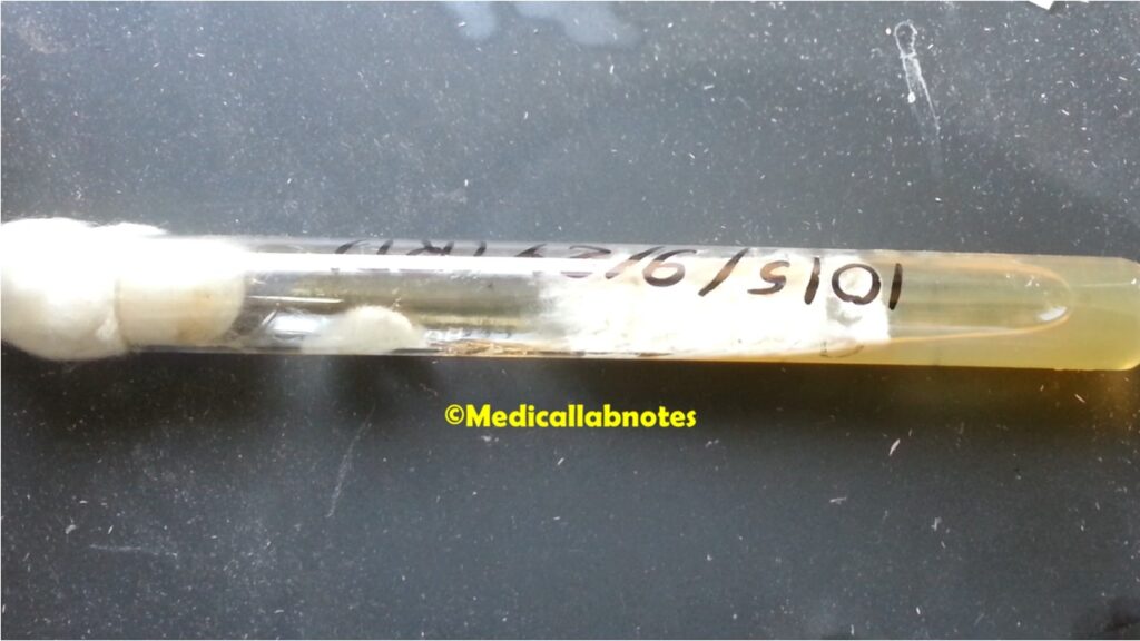
- Fungal Culture: Culturing the clinical specimen on appropriate fungal growth media is vital for the isolation and identification of Scedosporium. Sabouraud dextrose agar (SDA) is commonly used for this purpose. The cultures are incubated at appropriate temperatures (usually around 25-30°C) for several days to promote fungal growth.
- Macroscopic Examination: After incubation, the colonies of Scedosporium on the culture media can be examined macroscopically. The colonies are typically moist, creamy to beige in color, and may exhibit varying pigmentation depending on the species.
- Microscopic Examination of Pure Culture: Once the fungal colonies have grown, a slide culture can be prepared to observe the micro-morphological characteristics in detail. This may involve teasing apart the hyphae and conidia from the colony and mounting them on a slide for microscopic examination.
- Molecular Identification: Molecular techniques, such as DNA sequencing, are becoming increasingly important for accurate species identification of Scedosporium. PCR (polymerase chain reaction) assays targeting specific genetic markers can help differentiate between different species of Scedosporium and other related fungi.
- Antifungal Susceptibility Testing: Since they are known to be resistant to many antifungal drugs, antifungal susceptibility testing may be performed to determine the most effective treatment options. This involves testing the sensitivity of the isolated fungus to various antifungal agents.
- Histopathological Examination: In cases of invasive infections, histopathological examination of tissue biopsy specimens can reveal the presence of Scedosporium hyphae and conidia within affected tissues. This can provide valuable diagnostic information.

It’s important to note that working with Scedosporium in the laboratory requires appropriate biosafety precautions, as these fungi can pose a risk to laboratory personnel, especially in immunocompromised individuals.
Treatment
Treatment of Scedosporium infections can be challenging due to the intrinsic resistance of these fungi to many antifungal drugs. The choice of treatment depends on several factors, including the site and severity of infection, the immune status of the patient, and the species of Scedosporium involved. As with any medical condition, treatment decisions should be made in consultation with an infectious disease specialist or a healthcare professional experienced in managing fungal infections. Here are some general treatment considerations for Scedosporium infections:
- Antifungal Therapy: The mainstay of treatment for Scedosporium infections is antifungal medication. The choice of antifungal agent will depend on the species of Scedosporium and its susceptibility to specific drugs. Commonly used antifungal agents include voriconazole, posaconazole, itraconazole, and isavuconazole. In some cases, combination therapy with multiple antifungal agents may be necessary to improve treatment efficacy.
- High-Dose Therapy: Due to the resistance of Scedosporium to antifungal drugs, high doses of certain antifungal agents may be required to achieve therapeutic levels in the body.
- Duration of Treatment: The duration of antifungal therapy varies depending on the type and severity of the infection. Some infections may require prolonged treatment courses, even after clinical improvement, to prevent relapse.
- Monitoring: Close monitoring of the patient’s response to treatment and any adverse effects of antifungal medications is essential. Regular laboratory tests, imaging studies, and clinical assessments are typically conducted during treatment.
- Surgical Intervention: In some cases, surgical intervention may be necessary to remove infected tissues or abscesses, especially in cases of localized infections or when antifungal therapy alone is insufficient.
- Immune Support: In immunocompromised patients, managing the underlying condition and providing immune support can be crucial for a successful outcome. This may involve optimizing immunosuppressive therapy or administering therapies to boost the immune system.
- Antifungal Susceptibility Testing: In cases of difficult-to-treat infections or when standard therapies are not effective, antifungal susceptibility testing can guide treatment decisions. This helps identify the most effective antifungal agents against the specific strain of Scedosporium.
- Investigational Therapies: Since treatment options for Scedosporium infections are limited, participation in clinical trials of investigational antifungal agents may be considered in certain cases.
Prevention
Preventing Scedosporium infections is essential, especially in individuals who are immunocompromised or have underlying medical conditions that put them at higher risk of severe fungal infections. While it may not be possible to completely eliminate the risk, several preventive measures can be taken to reduce the likelihood of Scedosporium infections. Here are some strategies for prevention:
- Infection Control in Healthcare Settings: In hospitals and healthcare facilities, strict infection control practices should be implemented to minimize the spread of fungal pathogens, including Scedosporium. This includes proper hand hygiene, appropriate use of personal protective equipment, and adherence to isolation protocols for patients with suspected or confirmed fungal infections.
- Environmental Hygiene: They are commonly found in the environment, particularly in soil and water sources. Regular cleaning and maintenance of hospital environments and medical equipment can help reduce the exposure of vulnerable patients to the fungi.
- Monitoring and Surveillance: Regular monitoring and surveillance of patients at higher risk of fungal infections can help identify infections early and implement appropriate treatment promptly.
- Prophylactic Antifungal Therapy: In specific high-risk populations, such as organ transplant recipients or patients undergoing intensive chemotherapy, prophylactic antifungal therapy may be considered to prevent fungal infections, including those caused by Scedosporium.
- Immune Support: For individuals with compromised immune systems, measures to strengthen the immune system, such as vaccination and avoiding unnecessary immunosuppressive medications, can reduce the risk of fungal infections.
- Avoiding Environmental Exposure: While it may not be entirely avoidable, reducing exposure to environments with a higher concentration of Scedosporium, such as construction sites or areas with decaying organic matter, can lower the risk of infection.
- Educational Programs: Healthcare providers and patients should be educated about the risk factors for Scedosporium infections and the importance of early recognition and treatment. Patients with chronic lung conditions, such as cystic fibrosis, should be aware of the potential risks and practice good respiratory hygiene.
- Research and Development: Continued research on Scedosporium, including its ecology, pathogenicity, and antifungal resistance mechanisms, can lead to improved preventive measures and more effective treatments.
Keynotes
Scedosporium is a genus of medically important fungi with several key features and implications:
- Opportunistic Pathogens: They are opportunistic pathogens, causing infections primarily in individuals with compromised immune systems or underlying medical conditions.
- Wide Environmental Distribution: These fungi are commonly found in the environment, particularly in soil and water sources, making them ubiquitous worldwide.
- Clinical Presentations: Scedosporium infections can manifest in various ways, including respiratory infections, skin and soft tissue infections, and disseminated infections affecting multiple organs.
- Species Complexity: The genus includes several species, with S. apiospermum and S. prolificans being the most clinically significant. Accurate species identification is crucial for effective management.
- Resistance to Antifungal Drugs: They are intrinsically resistant to many antifungal agents, making treatment challenging and often requiring high doses or combination therapy.
- Biofilm Formation: Scedosporium can form biofilms, increasing its resilience to treatment and host immune defenses.
- Allergic Reactions: Exposure to Scedosporium can also lead to allergic reactions, especially in patients with pre-existing respiratory conditions.
- Laboratory Diagnosis: Diagnosis involves microscopic examination, fungal culture, molecular identification, and antifungal susceptibility testing.
- Treatment: Treatment options for Scedosporium infections are limited, and management usually involves antifungal therapy with close monitoring of patient response and possible surgical intervention.
- Prevention: Preventive measures focus on infection control in healthcare settings, environmental hygiene, and immune support for vulnerable individuals.
- Prognosis: Scedosporium infections can be severe, especially in immunocompromised patients, necessitating early diagnosis and prompt treatment.
- Research Importance: Ongoing research on Scedosporium is essential to improve understanding of its pathogenicity, antifungal resistance, and potential new treatment options.
Further Readings
- Lackner M, et al. (2014). Species recognition and clinical relevance of the zygomycetous genus Lichtheimia (syn. Absidia pro parte, Mycocladus). Journal of Clinical Microbiology, 52(4), 1156-1160.
- Cortez KJ, et al. (2008). Infections caused by Scedosporium spp. Clinical Microbiology Reviews, 21(1), 157-197.
- Biswas C, et al. (2018). Management of Scedosporium apiospermum central nervous system infections. Mycoses, 61(12), 917-926.
- Rodriguez-Tudela JL, et al. (2008). Susceptibility patterns in a large collection of Scedosporium and Pseudallescheria clinical isolates. Antimicrobial Agents and Chemotherapy, 52(6), 2123-2126.
- Steinbach WJ, et al. (2012). Breakthrough Scedosporium apiospermum infections in allogeneic hematopoietic stem cell transplant recipients receiving voriconazole. Clinical Infectious Diseases, 54(1), 161-169.
- Alastruey-Izquierdo A, et al. (2015). ECMM/ISHAM recommendations for clinical management of Scedosporium species and Lomentospora prolificans. Clinical Microbiology and Infection, 21(5), 403-408.
- Chakrabarti A, et al. (2014). Fungal infections in Asia: the current epidemiological scenario. Medical Mycology, 52(1), 13-23.
- Lackner M, et al. (2016). Scedosporium and Lomentospora: an updated overview of underrated opportunists. Medical Mycology, 54(4), 313-330.
- Blyth CC, et al. (2017). The impact of fungal co-infection in human immunodeficiency virus-infected individuals. Clinical Microbiology and Infection, 23(3), 216-223.
- Al-Hatmi AM, et al. (2016). Global molecular epidemiology and genetic diversity of Fusarium, a significant emerging group of human opportunists from 1958 to 2015. Emerging Microbes & Infections, 5(12), e124.
It¦s actually a great and useful piece of information. I¦m glad that you shared this useful info with us. Please keep us informed like this. Thanks for sharing.