
Introduction of Staphylococcus and Micrococcus
Table of Contents
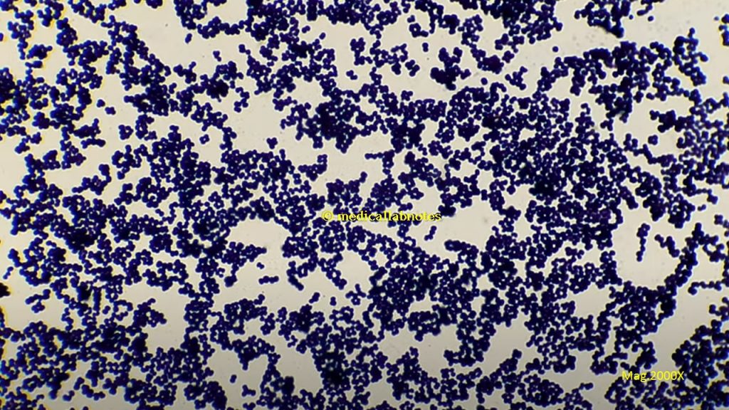
Staphylococcus (pleural-staphylococci) is spherical, non-motile, gram-positive in singles, pairs, and clusters. On nutrient agar, growth is opaque and golden yellow or white color. Catalase and coagulase test positive (Staphylococcus aureus), oxidase negative, aerobic or facultative anaerobe. Parasite of humans and animals. Normal flora of the skin, upper respiratory tract, and feces of humans, animals, and birds too. S. aureus is the etilogical agent of –

- Abscess
- Conjunctivitis
- Corneal ulcer
- Septicemia
- Endocarditis
- Pneumonia
- Mastitis: It is an inflammation of the breast.
- Empyema: It is an accumulation of pus in the body cavity.
- Food poisoning
- Staphylococcal Scalded Syndrome
- Toxic Shock Syndrome (TSS)-enterotoxin F
- Septic arthritis
- Meningitis
- Osteomyelitis
Species of Staphylococcus are-
- Staphylococcus aureus
- Staphylococcus schleriferi
- Staphylococcus felis
- Staphylococcus intermedius
- Staphylococcus lutrae
- Staphylococcus hyicus
- Staphylococcus lodgunensis
- Staphylococcus lodgunensis
- Staphylococcus schleriferi
- Staphylococcus hyicus
Micrococcus (pleural-micrococci) is free-living in the environment and also normal flora of the skin. It is Gram-positive cocci in tetrads, catalase-positive, coagulase-negative, arranged in clusters that differ from Staphylococcus in attacking sugars oxidatively which may appear in irregular clusters, groups of four or eight. It is often larger than Staphylococcus. Species of Micrococcus are-
- Micrococcus luteus
- Micrococcus terreus
- Micrococcus lylae
- Micrococcus mortus
- Micrococcus mucilaginosis
- Micrococcus roseus
Differences between Staphylococcus and Micrococcus
The differences between Staphylococcus and Micrococcus are as follows-
| Feature | Staphylococcus | Micrococcus |
| Anaerobic growth | Positive | Negative |
| Arrangement of cells (predominant) | Clusters | Clusters, tetrads |
| Clusters, tetrads | 30-39 | 64-75 |
| Carbohydrate utilization | Fermentative | Oxidative or nil |
| Oxidase (modified oxidase test) | Negative | Positive |
| 0.04U bacitracin | Resistant | Sensitive |
| Lysostaphin | Sensitive | Resistant |
| Teichoic acid in cell wall | Present | Absent |
Keynotes
- Staphylococcus is the Gram-positive cocci in clusters while Micrococcus is the Gram-positive cocci in tetrads.
- Staphylococcus aureus is the coagulase-positive (slide and tube both) whereas Micrococcus is modified oxidase-positive.
- Staphylococcus aureus generally expresses golden yellow color while Micrococcus luteus forms bright yellow colonies and Micrococcus roseus pink colonies on nutrient agar.
Staphylococcus and Micrococcus Footages
Gram-positive cocci in singles, pairs and cluster of Staphylococcus aureus in clinical sample, pus

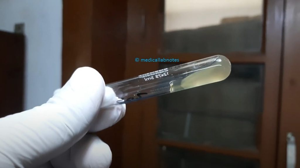
Staphylococcus aureus growth in Thioglycollate broth

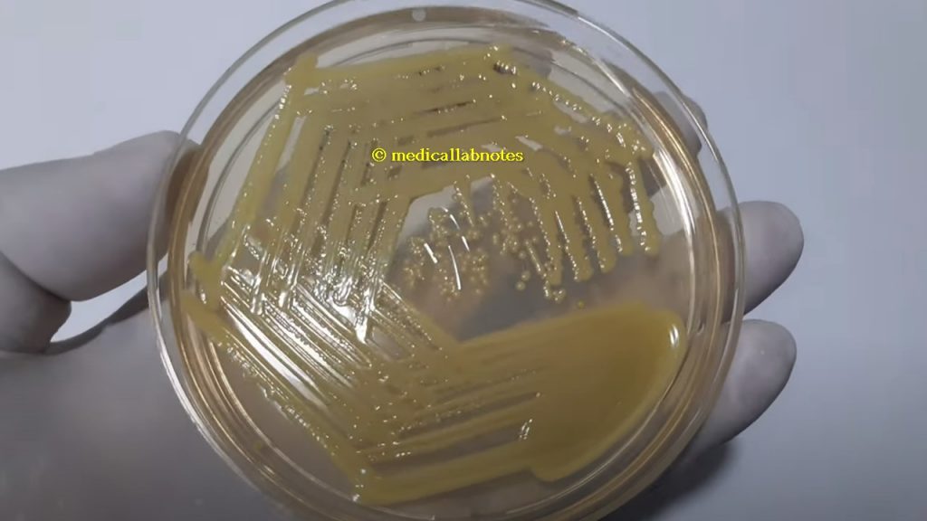
A yellow pigment staphyloxanthin producing strain of Staphylococcus aureus on nutrient agar

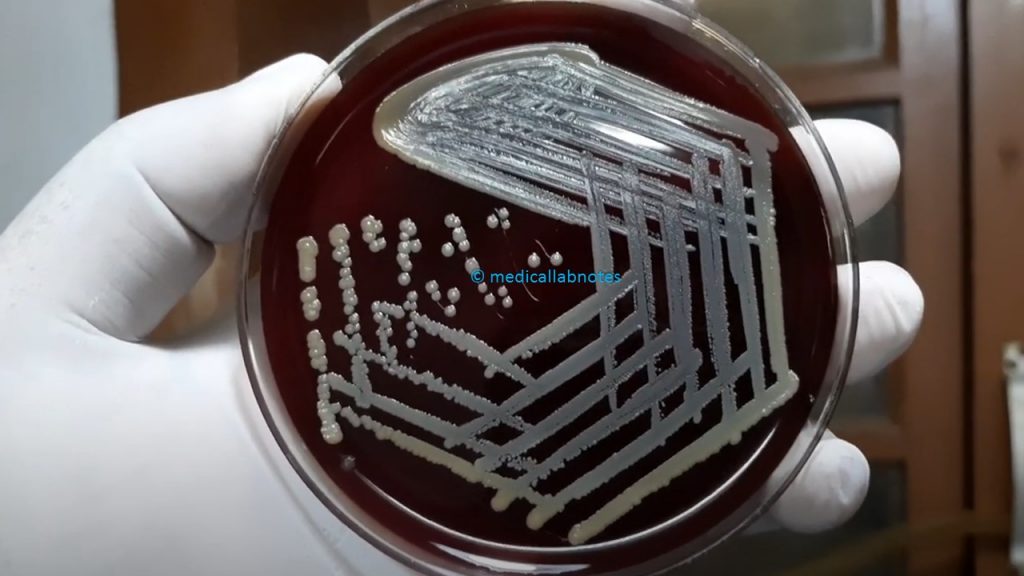
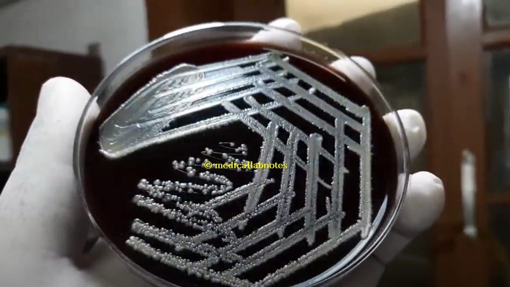
A golden yellow pigment producing strain of Staphylococcus aureus on blood agar
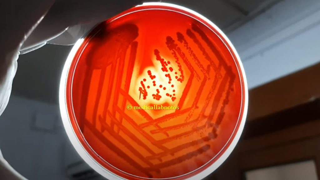
Beta-hemolytic colony of Staphylococcus aureus on blood agar demonstration
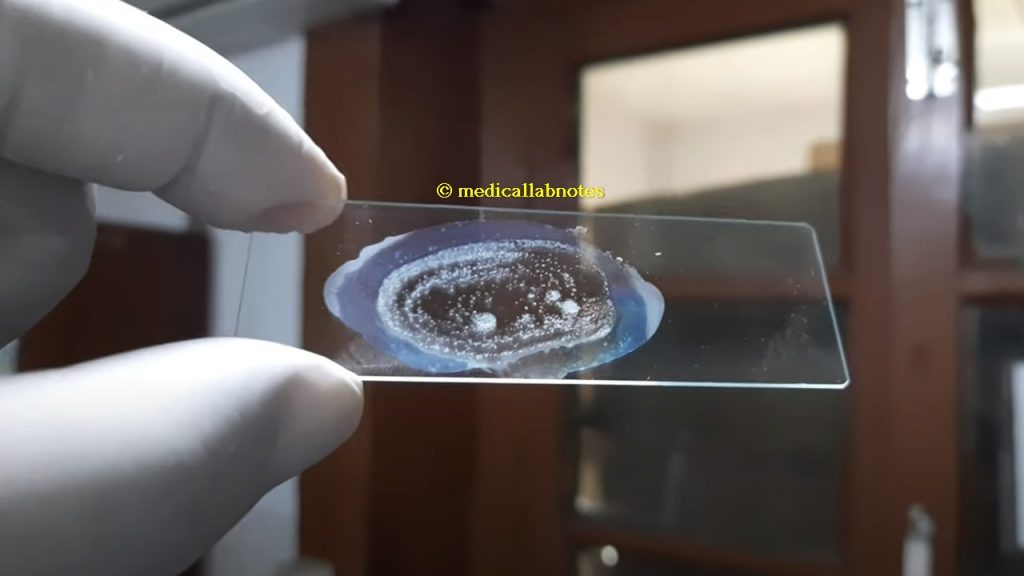
Slide coagulase positive Staphylococcus aureus
Tube coagulase positive Staphylococcus aureus
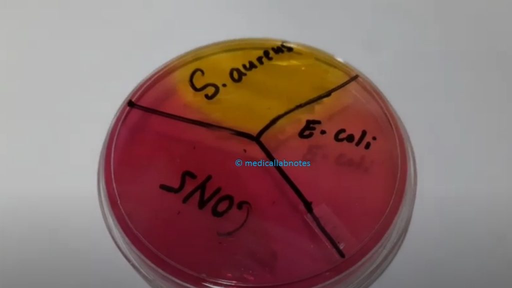
Staphylococcus aureus, CoNS and Escherichia coli growth on Mannitol Salt Agar ( MSA) Demonstration
Gram-positive cocci in singles, pairs and clusters or groups of Staphylococcus aureus in Gram staining of culture

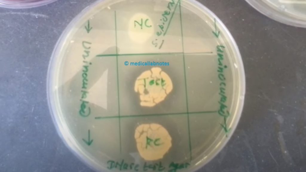
DNase Positive strain of Staphylococcus aureus
MRSA strain of Staphylococcus aureus on Muller-Hinton agar

D-Zone Test Positive Macrolide-lincosamide-streptogramin B (MLSB) strain of Staphylococcus aureus Demonstration

White colonies of Staphylococcus aureus on Muller-Hinton agar (MHA) Demonstration

Coagulase-negative staphylococci (CoNS) colony morphology on MSA

Coagulase-negative staphylococci (CoNS) colony characteristics on blood agar
Micrococcus roseus colony Morphology on blood agar

Micrococcus roseus Colony Morphology on Nutrient Agar

Micrococcus luteus Colony Morphology on Muller-Hinton Agar
Micrococcus in Gram staining of culture showing Gram-positive cocci in singles, pairs, tetrads and groups
Oxidase Positive Micrococcus luteus
Further Readings
- https://www.microbiologyresearch.org/docserver/fulltext/micro/30/3/mic-30-3-409.pdf?
- https://universe84a.com/collection/staphylococcus-versus-micrococcus/
- https://www.ncbi.nlm.nih.gov/pmc/articles/PMC271202/
- https://assets.publishing.service.gov.uk/government/uploads/system/uploads/attachment_data/file/832968/ID_7_dj_.pdf
- https://www.sciencedirect.com/science/article/pii/B9780121617752500207
- Bailey & Scott’s Diagnostic Microbiology. Editors: Bettey A. Forbes, Daniel F. Sahm & Alice S. Weissfeld, 12th ed 2007, Publisher Elsevier.
- Colour Atlas and Textbook of Diagnostic Microbiology. Editors: Koneman E.W., Allen D.D., Dowell V.R. Jr, and Sommers H.M.
- Cowan & Steel’s Manual for identification of Medical Bacteria. Editors: G.I. Barron & R.K. Felthani, 3rd ed 1993, Publisher Cambridge University Press.
- Jawetz, Melnick and Adelberg’s Medical Microbiology. Editors: Geo. F. Brook, Janet S. Butel & Stephen A. Morse, 21st ed 1998, Publisher Appleton & Lance, Co Stamford Connecticut.
- Mackie and Mc Cartney Practical Medical Microbiology. Editors: J.G. Colle, A.G. Fraser, B.P. Marmion, A. Simmous, 4th ed, Publisher Churchill Living Stone, New York, Melborne, Sans Franscisco 1996.
- Clinical Microbiology Procedure Handbook Vol. I & II, Chief in editor H.D. Isenberg, Albert Einstein College of Medicine, New York, Publisher ASM (American Society for Microbiology), Washington DC.
- Manual of Clinical Microbiology. Editors: P.R. Murray, E. J. Baron, M. A. Pfaller, F. C. Tenover and R. H. Yolken, 7th ed 2005, Publisher ASM, USA
- District Laboratory Practice in Tropical Countries – Part-2- Monica Cheesebrough- 2nd Edn Update
- Textbook of Diagnostic Microbiology. Editors: Connie R. Mahon, Donald G. Lehman & George Manuselis, 3rd edition2007, Publisher Elsevier.
- Topley & Wilsons Principle of Bacteriology, Virology, and immunology Vol I, II, III, IV & V. Editors: M.T. Parker & L.H. Collier, 8th ed 1990, Publisher Edward Arnold publication, London.
- Medical Microbiology-The Practice of Medical Microbiology Vol-2-12th Edn. –Robert Cruickshank
I loved as much as you’ll receive carried out right here. The sketch is attractive, your authored subject matter stylish. nonetheless, you command get bought an nervousness over that you wish be delivering the following. unwell unquestionably come more formerly again as exactly the same nearly a lot often inside case you shield this increase.
Really great visual appeal on this site, I’d rate it 10 10.
I truly appreciate your work, Great post.
I consider something really interesting about your web site so I saved to my bookmarks.
I was looking at some of your content on this internet site and I believe this internet site is rattling informative ! Keep on putting up.
Hello, I think your blog might be having browser compatibility issues. When I look at your website in Safari, it looks fine but when opening in Internet Explorer, it has some overlapping. I just wanted to give you a quick heads up! Other then that, excellent blog!
Gοod post. I learn something totally new and challenging on blogs I stumbleuρon on a daily basis.
It will always be exⅽiting to read artticles from other
authors and practice something from other sites.
It’s actually a great and useful piece of information. I am glad that you just shared this helpful info with us. Please stay us informed like this. Thank you for sharing.
Great wordpress blog here.. It’s hard to find quality writing like yours these days. I really appreciate people like you! take care
I also conceive thus, perfectly indited post! .
I really like your writing style, fantastic information, regards for putting up :D. “All words are pegs to hang ideas on.” by Henry Ward Beecher.
I enjoy assembling utile information , this post has got me even more info! .
Thanks for sharing, this is a fantastic article post. Awesome.
Have you ever considered writing an e-book or guest authoring on other sites? I have a blog based on the same ideas you discuss and would really like to have you share some stories/information. I know my visitors would enjoy your work. If you’re even remotely interested, feel free to shoot me an email.
Wow, great post.Really looking forward to read more. Want more.
I really enjoy the post.Really looking forward to read more. Cool.
I value the blog post. Really Cool.
Looking forward to reading more. Great article post. Really Great.
I’m often to blogging and i in actual fact respect your content. The piece has actually peaks my interest. I’m going to bookmark your content and preserve checking for brand new information.
Very good blog.Really looking forward to read more. Awesome.
Great article post. Much obliged.
Very neat blog post.Really looking forward to read more. Really Cool.
Looking forward to reading more. Great article post.Really looking forward to read more. Fantastic.
Everyone loves what you guys are up too. This kind of clever work and exposure! Keep up the wonderful works guys I’ve included you guys to blogroll.
Thanks-a-mundo for the article post.Much thanks again. Great.
side effects for sulfamethoxazole trimethoprim keflex
Looking forward to reading more. Great blog post.Much thanks again. Fantastic.
I loved your article post.Really looking forward to read more. Will read on…
Im thankful for the post.Really thank you! Fantastic.
Good post however I was wanting to know if you could write a litte more on this topic? I’d be very thankful if you could elaborate a little bit more. Cheers!
Looking forward to reading more. Great blog post.Much thanks again. Great.
Fantastic blog. Keep writing.
Heya i’m for the first time here. I foundthis board and I find It truly useful & it helped me out a lot.I hope to give something back and aid others like you aided me.
Keep up the fantastic work, I read few content on this site and I believe that your site is very interesting and contains bands of great information.
Thanks a lot for the article post.Much thanks again. Cool.
Hello there, I discovered your site by the use of Google at the same time as searching for a similar topic, your website came up, it seems to be good. I’ve bookmarked it in my google bookmarks.