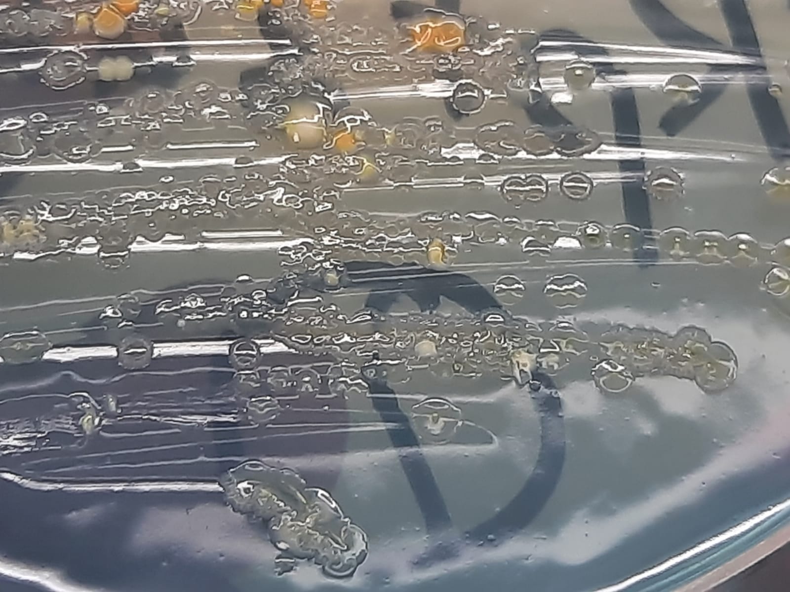
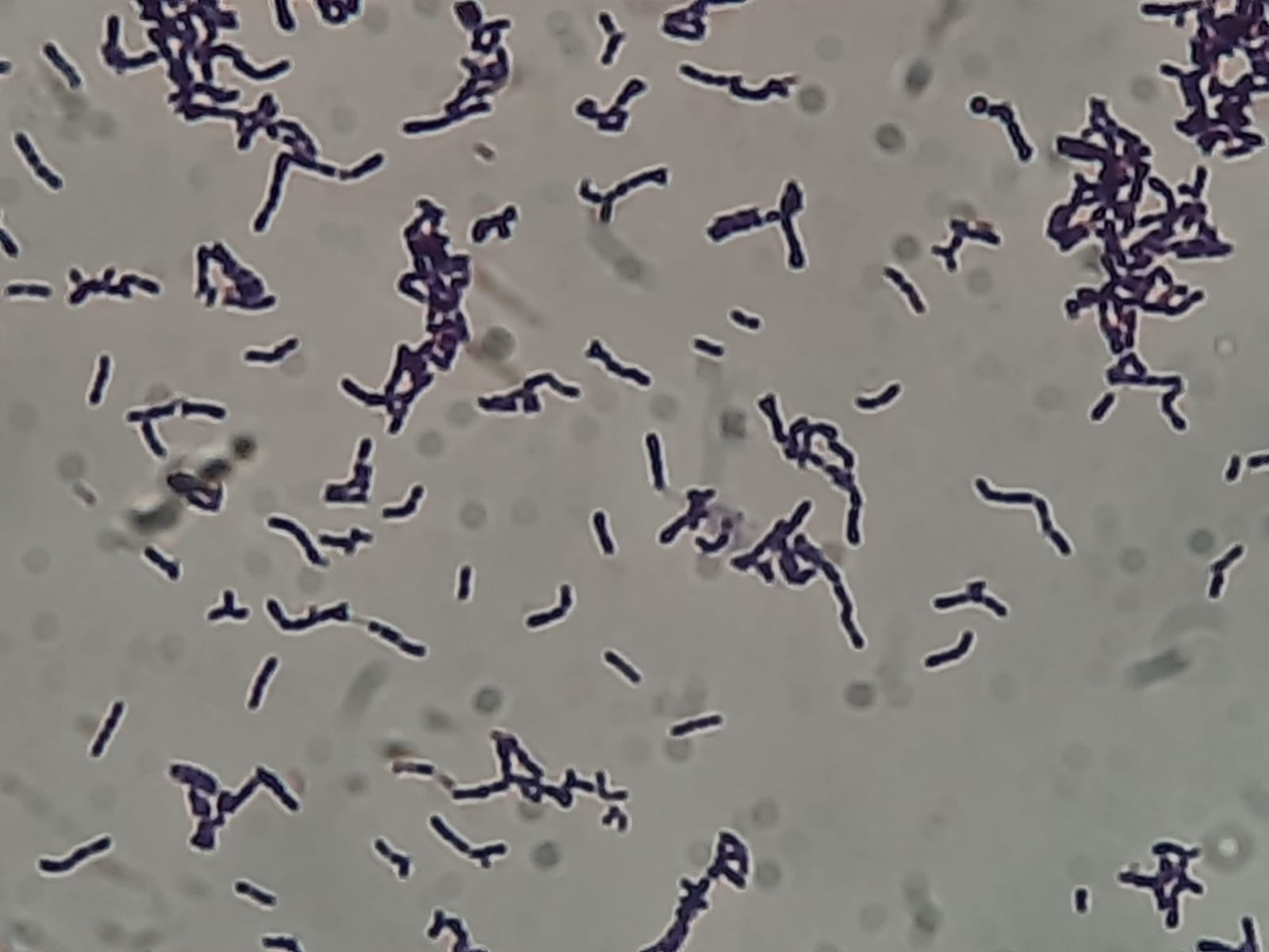
Contamination in L-J Media: Introduction, Common Contaminant, Identification clues, Minimization tricks, and Keynotes 

Advanced Fungal Contamination and Decolorization of L-J Medium: Fungal Overgrowth-The slants show dense, white, "cotton-like" mycelial growth. This morphology is a classic indicator of contamination by environmental molds, such as Aspergillus or Penicillium. Media Alteration- There is a total loss of the characteristic malachite green color. The medium has turned pale yellow or colorless, suggesting a significant shift in pH or the enzymatic degradation of the selective agent by the contaminants. Diagnostic Interpretation-The rapid growth of these organisms has completely masked any potential slow-growing Mycobacterium tuberculosis. These cultures are invalid and must be recorded as contaminated.

Introduction Lowenstein-Jensen (L-J) medium is the gold standard for the cultivation of Mycobacterium tuberculosis. However, because it is an egg-based, non-selective (or semi-selective) medium that requires long incubation periods, it is highly susceptible to contamination. L-J medium is rich in nutrients (eggs, glycerol, potato flour), …

Read more

Acinetobacter haemolyticus-Introduction, Morphology, Pathogenicity, Lab Diagnosis, Treatment, Prevention, and Keynotes

Acinetobacter haemolyticus growth on CLED agar

Introduction Acinetobacter haemolyticus is an aerobic, non-fermenting bacterium found in nature and hospital environments. It belongs to the Acinetobacter genus, frequently causing healthcare-associated infections. It is generally considered less virulent than Acinetobacter baumannii but is increasingly recognized in human infections.  Morphology Pathogenicity Lab Diagnosis Treatment Prevention Keynotes Further Readings

HiCrome Candida Differential Agar: Introduction, Principle, Test Requirement, Procedure, Result-Interpretation, and Keynotes

Various Candida species growth on HiCrome Candida Differential Agar after 4 days of incubation

Introduction HiCrome™ Candida Differential Agar is a specialized chromogenic medium used in clinical microbiology for the rapid isolation and presumptive identification of Candida species. The medium is designed to differentiate between major Candida species—Candida albicans, Candida tropicalis, Candida krusei (Pichia kudriavzevii), and Candida glabrata—based on colony color and morphology within 48 hours. …

Read more

Rothia kristinae: Introduction, Pathogenicity, Lab Diagnosis, Treatment, Prevention, and Keynotes

R. kristinae in Gram-stained culture microscopy shows irregular, pleomorphic Gram-positive bacilli, ranging from short rods to e

Introduction Pathogenicity While typically considered a low-grade commensal, it causes serious infections primarily in immunocompromised patients or those with indwelling medical devices.  Lab Diagnosis Treatment There are no official clinical breakpoints, so treatment is often guided by expert consultation and raw Minimum Inhibitory Concentration (MIC) values.  Prevention Keynotes …

Read more

Serpentine Cord: Introdction, Clinical Significance, and Keynotes

Serpentine cord or Cord factor or Trehalose dimycolate,TDM of Mycobacterium tuberculosis complex (MTBC) in Ziehl-Neelsen Staining of modified Middlebrook 7H9 Broth at a magnification of 1600X

Introduction Clinical Significance Keynotes Further Readings

Fungal Culture of Clinical Samples: Introduction, Principle, Clinical Significance, and Keynotes

Body fluid and sputum for culture

Introduction Fungal culture is the gold standard method for the detection and identification of pathogenic fungi in clinical microbiology. It is essential for diagnosing superficial, subcutaneous, and systemic mycoses. Clinical specimens such as blood, sputum, bronchoalveolar lavage (BAL), urine, cerebrospinal fluid (CSF), skin scrapings, nail …

Read more

Fungal Identification by Artificial Intelligence (AI): Introduction, Working Mechanisms, Clinical Significance, and Keynotes

Yeast and mold growth on SDA agar

Introduction Fungal infections represent a growing concern in both immunocompromised and immunocompetent individuals. Conventional identification methods, including culture, microscopy, and biochemical testing, often require several days and may lack sensitivity or specificity for certain opportunistic fungi. Molecular techniques such as PCR and sequencing have improved …

Read more

Comparative Microscopy of Candida Species: Introduction, Table, and Keynotes

Chlamydospores of Candida albicans found in direct clinical sample(sputum) KOH mount Microscopy (Mag.1600X)

Introduction The genus Candida comprises a diverse group of opportunistic yeasts frequently isolated from human clinical specimens. Among them, Candida albicans remains the most common cause of infections; however, non-albicans Candida species, such as C. tropicalis, C. glabrata, and C. krusei, are increasingly recognized as important pathogens in immunocompromised patients, …

Read more