
Introduction
Table of Contents
Trichophyton mentagrophytes is a dermatophyte fungus belonging to the genus Trichophyton, which is known for causing various skin infections in humans and animals. It is an anthropophilic fungus, meaning it primarily infects humans, but it can also infect animals like rodents and domesticated pets. The term “dermatophyte” refers to a group of fungi that have the ability to invade and grow on the keratinized tissues of the skin, hair, and nails.
Skin infections caused by Trichophyton mentagrophytes are commonly referred to as dermatophytosis or “ringworm,” although the condition is not caused by a worm but by the fungal infection. The name “ringworm” comes from the classic red, circular rash that appears on the skin, resembling a worm’s shape.
Transmission of Trichophyton mentagrophytes occurs through direct contact with infected individuals, animals, or contaminated objects, such as towels, clothing, or shared surfaces in public places like gyms or swimming pools. The fungus thrives in warm and humid environments, making these places ideal for its growth and transmission.
Typical symptoms of Trichophyton mentagrophytes infection include red, itchy, and scaly patches on the skin that may be accompanied by raised edges, giving them a ring-like appearance. The condition can affect various areas of the body, including the scalp, feet (athlete’s foot), groin (jock itch), and other body parts.
Diagnosis is usually made through clinical examination, microscopic examination of skin scrapings, or fungal cultures to identify the specific species of fungus involved. Treatment often involves topical antifungal medications for mild cases and oral antifungal medications for more severe or widespread infections.
Prevention involves maintaining good personal hygiene, avoiding sharing personal items with infected individuals, and keeping the skin clean and dry, especially in areas prone to moisture accumulation.
It is important to consult a healthcare professional if you suspect you have a fungal skin infection to receive appropriate diagnosis and treatment. Fungal infections can be persistent, so early intervention is essential to prevent the spread of the infection and to facilitate a faster recovery.
Morphology
Trichophyton mentagrophytes exhibits distinctive morphology that aids in its identification under the microscope and in culture. As a dermatophyte fungus, its morphology is well-adapted for invading and colonizing keratinized tissues like the skin, hair, and nails. Here are some key characteristics of Trichophyton mentagrophytes:
- Macroscopic appearance on culture:
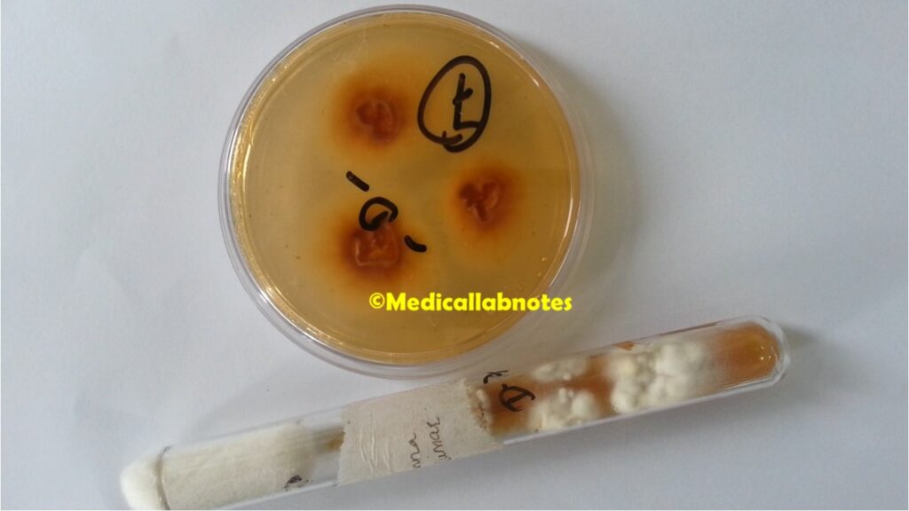
- Colonies on culture media typically appear woolly or cottony with a white to cream coloration. As they mature, the color may change to yellow or tan.
- The colony growth rate is relatively fast, usually reaching a diameter of a few centimeters within a week.
- Microscopic appearance under the microscope:
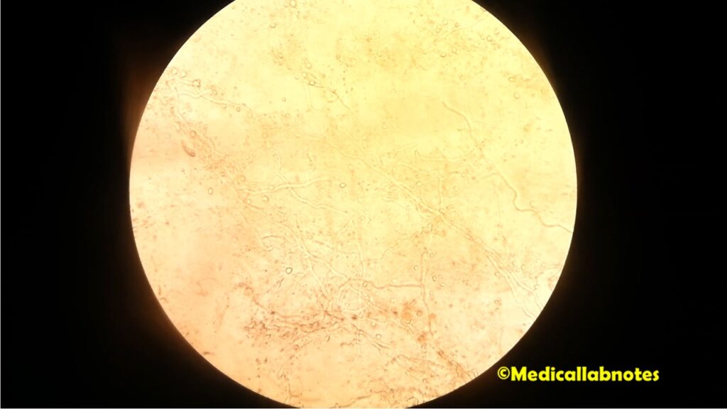
- Trichophyton mentagrophytes produces septate hyphae, meaning the hyphae are divided into compartments by cross-walls called septa. These hyphae are the branching structures that make up the body of the fungus.
- The hyphae of Trichophyton mentagrophytes can be both smooth-walled and rough-walled (rugose), which is a useful distinguishing feature in differentiating it from other species.
- It forms numerous spindle-shaped or club-shaped macroconidia (asexual spores) that are usually found in clusters along the hyphae. These macroconidia are multicellular and have thick walls with characteristic rough surfaces, often described as “lumpy” or “knobby.”
- Microconidia (smaller asexual spores) are usually present as well, but they are less common and are typically not as numerous as the macroconidia.
- Terminal chlamydospores may also be present, which are thick-walled, rounded structures formed at the tips of hyphae and play a role in survival and persistence.
It is important to note that the morphology of Trichophyton mentagrophytes can vary slightly depending on the environmental conditions and the specific strain of the fungus. To accurately identify the species, cultures may be grown on specific media and observed under the microscope for characteristic features.
Pathogenicity
Trichophyton mentagrophytes is considered a highly pathogenic dermatophyte fungus, meaning it has the ability to cause a wide range of infections in humans and animals. Its pathogenicity is primarily due to its ability to invade and colonize keratinized tissues such as the skin, hair, and nails. The fungus secretes enzymes that can break down keratin, the tough protein that forms the structural basis of these tissues, allowing it to obtain nutrients and proliferate.
The main clinical manifestations of Trichophyton mentagrophytes infections include:
- Tinea corporis (ringworm of the body): This infection appears as red, scaly, and ring-shaped patches on the skin, often with raised edges. The central area of the rash may clear, giving it the characteristic ring-like appearance.
- Tinea pedis (athlete’s foot): A common infection affecting the feet, particularly between the toes and on the soles. It causes itching, burning, redness, and scaling.
- Tinea cruris (jock itch): This affects the groin area and is more common in males. It presents as red, itchy, and scaly patches in the groin and inner thighs.
- Tinea capitis (scalp ringworm): Primarily affects children and can lead to hair loss, scaling, and the formation of black dots (broken-off hair shafts) on the scalp.
- Tinea unguium (onychomycosis): Infection of the nails, causing them to become discolored, thickened, brittle, and distorted.
Trichophyton mentagrophytes is also zoonotic, meaning it can be transmitted between animals and humans. It is known to cause ringworm in domesticated pets, such as dogs and cats, and can be transmitted from infected animals to humans and vice versa.
The transmission of Trichophyton mentagrophytes occurs through direct contact with infected individuals, animals, or contaminated objects. It thrives in warm and humid environments, making places like public pools, locker rooms, and communal bathing areas common sources of infection.
Treatment of Trichophyton mentagrophytes infections typically involves topical or oral antifungal medications, depending on the severity and location of the infection. Early diagnosis and appropriate treatment are essential to prevent the spread of the infection to others and to avoid complications, such as secondary bacterial infections or chronic infections.
Lab Diagnosis
The laboratory diagnosis of Trichophyton mentagrophytes infection involves the examination of clinical samples collected from the affected area. The most common specimens used for diagnosis include skin scrapings, nail clippings, or hair samples. The diagnosis is primarily based on direct microscopic examination and culture of the collected samples. Here’s an overview of the lab diagnosis process:
- Direct Microscopic Examination:
- The collected sample (skin scrapings, nail clippings, or hair) is placed on a glass slide and treated with a drop of potassium hydroxide (KOH) solution.
- The KOH helps to dissolve the non-fungal elements, such as skin debris and keratin, making the fungal elements more visible under the microscope.
- The slide is then examined under a microscope at various magnifications to observe the fungal structures, including hyphae, macroconidia, and microconidia characteristic of Trichophyton mentagrophytes.
- Fungal Culture:
- A portion of the clinical sample is inoculated onto appropriate culture media that support the growth of dermatophytes.
- Sabouraud dextrose agar with cycloheximide and chloramphenicol is commonly used for fungal cultures.
- The culture plates are then incubated at the appropriate temperature (usually around 25-30°C) for up to several weeks, as dermatophytes typically grow slowly.
- The presence of Trichophyton mentagrophytes is confirmed by the characteristic appearance of the colony, which may include features such as texture, color, and rate of growth.
- Identification of Trichophyton mentagrophytes:
- Once the growth is observed on the culture plate, further identification tests may be performed to confirm the species.
- These tests may include microscopic examination of the fungal structures, such as macroconidia and microconidia, to distinguish Trichophyton species from other dermatophytes.
- Molecular methods like polymerase chain reaction (PCR) and DNA sequencing may also be used for accurate identification, especially in cases where differentiation between closely related species is required.

Treatment
The treatment of Trichophyton mentagrophytes infections, like other dermatophytosis (ringworm) infections, typically involves antifungal therapy. The choice of treatment depends on the severity of the infection, the affected area of the body, and the patient’s medical history. Treatment options include topical and oral antifungal medications. In some cases, a combination of both may be prescribed. It’s important to note that treatment should be continued until the infection has completely cleared, which may take several weeks. Here are the common treatment approaches:
- Topical Antifungal Medications:
- Clotrimazole: Available in creams, lotions, or powders, clotrimazole is effective against Trichophyton mentagrophytes and other dermatophytes. Apply the topical medication directly to the affected area, and the surrounding skin, following the instructions provided by your healthcare provider.
- Miconazole: Similar to clotrimazole, miconazole is available in various topical formulations and works effectively against Trichophyton mentagrophytes. Follow your doctor’s instructions on how to use the medication properly.
- Terbinafine: Topical terbinafine formulations are also effective against Trichophyton mentagrophytes. They are available as creams or gels and are generally applied once or twice daily.
- Oral Antifungal Medications:
- Griseofulvin: In cases of severe or widespread infections, oral antifungal medications like griseofulvin may be prescribed. Griseofulvin works systemically to inhibit fungal growth. It is usually taken daily for several weeks to months, depending on the location and extent of the infection.
- Terbinafine: Besides topical formulations, terbinafine is also available in oral tablets. It is generally well-tolerated and can be an effective treatment option for Trichophyton mentagrophytes infections.
- Combination Therapy:
- In some cases, especially when dealing with chronic or difficult-to-treat infections, healthcare providers may recommend a combination of topical and oral antifungal medications to enhance effectiveness and hasten healing.
Remember to follow your healthcare provider’s instructions carefully, including the duration and frequency of medication use. It’s crucial to complete the full course of treatment even if the symptoms improve, as stopping early may lead to recurrence or the development of drug-resistant strains.
Additionally, to aid in the recovery process and prevent reinfection, consider the following measures:
- Keep the affected area clean and dry.
- Avoid sharing personal items like towels, clothing, and combs with others.
- Wash and dry clothing and bedding regularly to prevent the spread of the fungus.
- Wear clean, breathable socks and well-ventilated footwear, especially in cases of athlete’s foot.
If the infection does not improve with the recommended treatment or if it worsens, it’s essential to consult your healthcare provider for further evaluation and adjustments to the treatment plan.
Prevention
Preventing Trichophyton mentagrophytes infections, also known as dermatophytosis or ringworm, involves adopting good personal hygiene practices and avoiding contact with infected individuals or contaminated objects. Here are some preventive measures to reduce the risk of acquiring or spreading Trichophyton mentagrophytes infections:
- Maintain Good Hygiene:
- Regularly wash and thoroughly dry your skin, especially in areas that are prone to sweating, such as the feet, groin, and underarms.
- Use soap and warm water to clean your body and hands, paying attention to areas between the fingers and toes.
- Dry yourself completely, including in between skin folds, after bathing or swimming.
- Keep Skin and Nails Dry:
- Moist environments provide a favorable breeding ground for dermatophytes. Avoid prolonged exposure to wet and humid conditions.
- After swimming or exercising, change into dry clothes promptly.
- For individuals with sweaty feet, consider using talcum powder or antifungal powder to keep the feet dry.
- Avoid Sharing Personal Items:
- Do not share towels, clothing, socks, shoes, combs, hairbrushes, or other personal items with others, especially if you suspect they may have a fungal infection.
- Wash clothing and towels with hot water and detergent after each use, especially if they have come into contact with an infected person.
- Protect Feet in Public Areas:
- Wear flip-flops or sandals in communal showers, locker rooms, and public swimming pools to reduce the risk of picking up the fungus from contaminated surfaces.
- Use Proper Footwear:
- Choose shoes that allow proper ventilation and avoid tight-fitting or non-breathable footwear, as they can create a warm and moist environment ideal for fungal growth.
- Maintain Pet Hygiene:
- If you have pets, regularly inspect them for signs of skin infections, especially if they spend time outdoors or interact with other animals.
- Consult a veterinarian if you suspect your pet may have a fungal infection to prevent transmission to humans and other animals.
- Practice Good Nail Hygiene:
- Keep your nails clean, dry, and trimmed. Avoid sharing nail clippers and other nail care tools.
- Educate Yourself and Others:
- Learn about the symptoms and modes of transmission of dermatophyte infections. Educate family members and close contacts to ensure everyone takes appropriate precautions.
- Promptly Treat Infections:
- If you suspect you have a fungal skin infection, seek medical attention promptly for diagnosis and treatment. Early intervention can help prevent the infection from spreading and becoming more difficult to treat.
Keynotes
Here are some keynotes on Trichophyton mentagrophytes:
- Fungal Pathogen: Trichophyton mentagrophytes is a dermatophyte fungus belonging to the genus Trichophyton, known for causing dermatophytosis (ringworm) infections in humans and animals.
- Anthropophilic and Zoophilic: It is primarily an anthropophilic fungus, meaning it primarily infects humans. However, it can also infect animals like rodents and domesticated pets, making it a zoonotic fungus.
- Mode of Transmission: The fungus is transmitted through direct contact with infected individuals, animals, or contaminated objects. It thrives in warm and humid environments, making public places like gyms, swimming pools, and communal bathing areas common sources of infection.
- Clinical Manifestations: Trichophyton mentagrophytes can cause various skin infections, including tinea corporis (ringworm of the body), tinea pedis (athlete’s foot), tinea cruris (jock itch), tinea capitis (scalp ringworm), and tinea unguium (onychomycosis).
- Morphology: Under the microscope, Trichophyton mentagrophytes exhibits septate hyphae with smooth and rough-walled (rugose) characteristics. It forms numerous spindle-shaped or club-shaped macroconidia with rough surfaces and terminal chlamydospores.
- Treatment: The treatment of Trichophyton mentagrophytes infections involves topical and/or oral antifungal medications. Topical antifungals like clotrimazole, miconazole, and terbinafine are used for milder infections, while oral antifungals like griseofulvin and terbinafine are prescribed for severe or widespread infections.
- Prevention: Preventive measures include maintaining good personal hygiene, keeping the skin and nails dry, avoiding sharing personal items, using proper footwear in public areas, and promptly treating infections to prevent spread.
- Zoonotic Potential: Trichophyton mentagrophytes can infect animals, and pet owners should be cautious and seek veterinary care if their pets show signs of skin infections.
- Contagiousness: The fungus is contagious and can spread easily, especially in shared living spaces, schools, and other crowded areas.
- Diagnosis: Diagnosis is typically made through clinical examination, microscopic examination of skin scrapings, and fungal cultures to identify the specific species involved.
Further Readings
- PubMed: PubMed is a free database maintained by the National Library of Medicine (NLM) that provides access to a vast collection of biomedical literature, including research articles and reviews on Trichophyton mentagrophytes. You can search for specific topics or keywords related to this fungus to find relevant studies and publications.
- Clinical Dermatology, 6th Edition: This comprehensive textbook by Thomas P. Habif covers various aspects of dermatology, including fungal skin infections. It may have dedicated chapters or sections on dermatophytes like Trichophyton mentagrophytes.
- Molecular Typing in Dermatophytes: This book, edited by Anil Kumar and published in 2018, focuses on molecular methods used for the identification and characterization of dermatophytes, including Trichophyton species.
- Journal of Medical Microbiology: This journal often publishes articles related to fungal infections, including studies on Trichophyton mentagrophytes and its epidemiology, pathogenesis, diagnosis, and treatment.
- Mycoses: Mycoses is a medical journal that covers research on fungal infections, including dermatophytosis caused by Trichophyton species.
- Textbook of Dermatology, Venereology, and Leprology, 3rd Edition: This textbook, edited by R. G. Valia and Ameet N. Valia, may contain valuable information on dermatophyte infections.
- European Journal of Clinical Microbiology & Infectious Diseases: This journal publishes articles on infectious diseases, including studies on the clinical aspects and antifungal resistance of Trichophyton species.