Direct Microscopy-KOH Smear: Introduction, Uses, and Keynotes
Introduction
Table of Contents
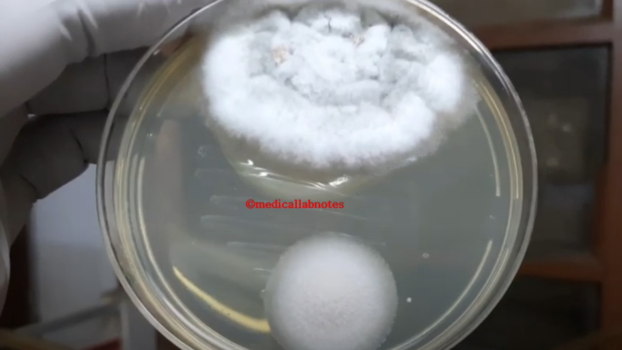
Direct microscopic examination is one of the most rapid and inexpensive diagnostic methods for detecting fungal elements in clinical specimens. Potassium hydroxide (KOH) smear is a routine technique in mycology that helps visualize fungal structures such as hyphae, yeast cells, and sclerotic bodies directly from patient samples. It is widely used as a first-line screening test for suspected fungal infections before culture or molecular testing.
Uses of Direct Microscopy-KOH Smear
Rapid Screening – Provides immediate results within minutes, aiding in early diagnosis.
Fig. Melanized septate fungal hyphae in KOH mount of BAL sampleFig. Yeast cells and budding yeasts in a KOH mount of Urine
Fig. Yeast cells, budding yeasts, and pseudohyphae in KOH mount of sputum, microscopic examination at a magnification of 1600XFig. Yeast cells and budding yeasts in KOH mount of urinary sediment microscopy at a magnification of 1600XFig. Septate acute-angled branching hyphae of Aspergillus in a KOH mount of sputumfIG. Fungal elements in the KOH mount of the urine sample at a magnification of 1600XFig. Chlamydospores of Candida albicans and hyphae in KOH mount of sputum microscopy at a magnification of 1600XFig. Heavy load of fungal elements in KOH mount of sputum specimen (Mag. 1600X)
Fig. In a potassium hydroxide (KOH) skin scraping for chromoblastomycosis, a sclerotic body is a characteristic, dark-brown, thick-walled, round, and multicellular fungal cell that appears as a “medlar body” or “muriform cell,” as shown in this image.