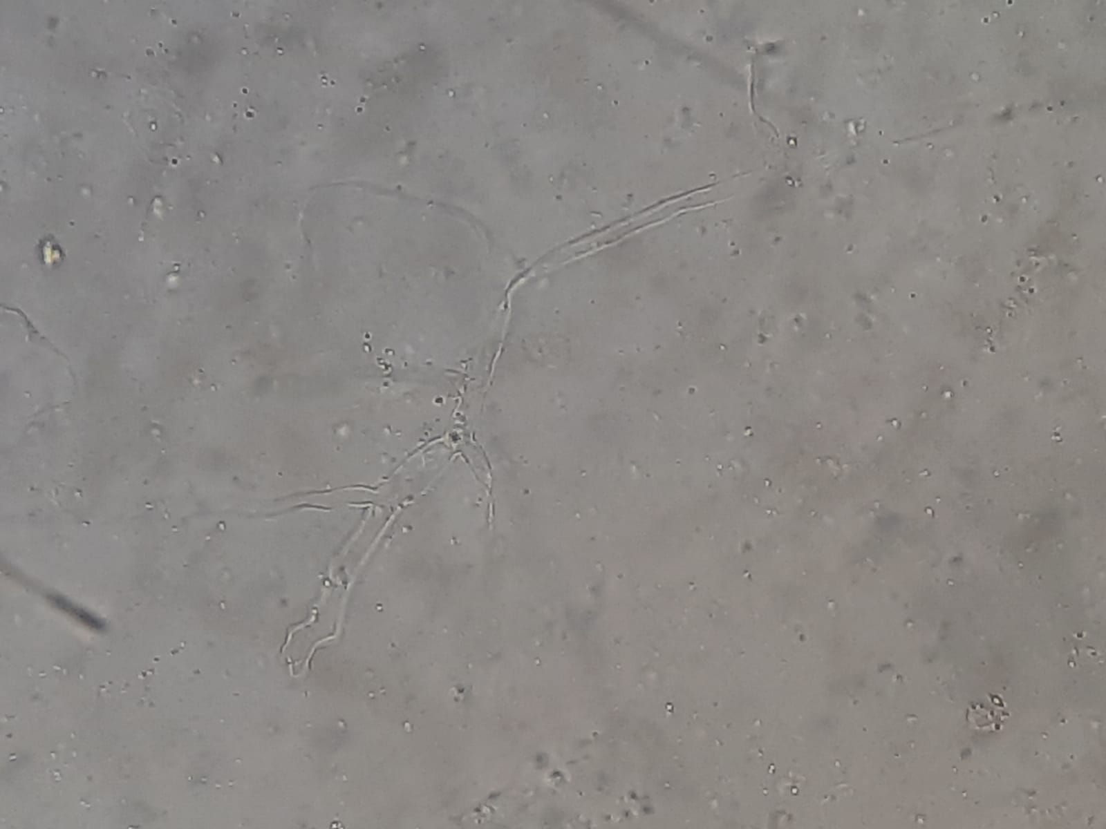
Fungal Elements in KOH Mount of Sputum Microscopy at a magnification of 1600X
Table of Contents
KOH mount of sputum is a rapid, direct microscopic technique used to detect fungal elements in the respiratory tract.
A 10–20% Potassium Hydroxide (KOH) solution digests mucus, epithelial cells, and debris in sputum, while preserving the chitin-rich fungal cell walls, making them appear clear, refractile, and easily visible.
This test is important for diagnosing pulmonary fungal infections, especially in patients with:
It rapidly identifies fungi such as Candida, Aspergillus, Mucorales, and Fusarium
Immediate bedside/bench-side detection of fungal elements before culture or molecular testing.
Useful for identifying:
Essential in ICU, diabetic, oncology, and transplant patients suspected of fungal pneumonia.
Quick detection of hyphae allows clinicians to start urgent antifungal therapy, especially in mucormycosis and invasive aspergillosis.
Correlates with:
Helps detect fungal infections co-existing with tuberculosis or HIV.
Introduction Classification: Belongs to the family Enterobacteriaceae. Habitat: Widely found in soil, water, plants, and…
Introduction Streptococcus mitis is a Gram-positive, alpha-hemolytic bacterium commonly found as a harmless commensal in…
Introduction An anemometer measures wind speed. It also measures wind pressure. The name comes from…
Introduction TB-LAMP (Tuberculosis Loop-Mediated Isothermal Amplification) is a manual, rapid molecular diagnostic test endorsed by…
Introduction The NALC-NaOH (N-acetyl-L-cysteine–sodium hydroxide) method is the gold standard for processing clinical specimens in…
Introduction The BD BACTEC™ MGIT™ 960 (Mycobacteria Growth Indicator Tube) is a fully automated, high-volume…