Pleural Fluid Microscopy: Introduction, Principle, Test Requirements, Procedure, Finding, Clinical Significance, and Keynotes

Collected Pleural Fluid-

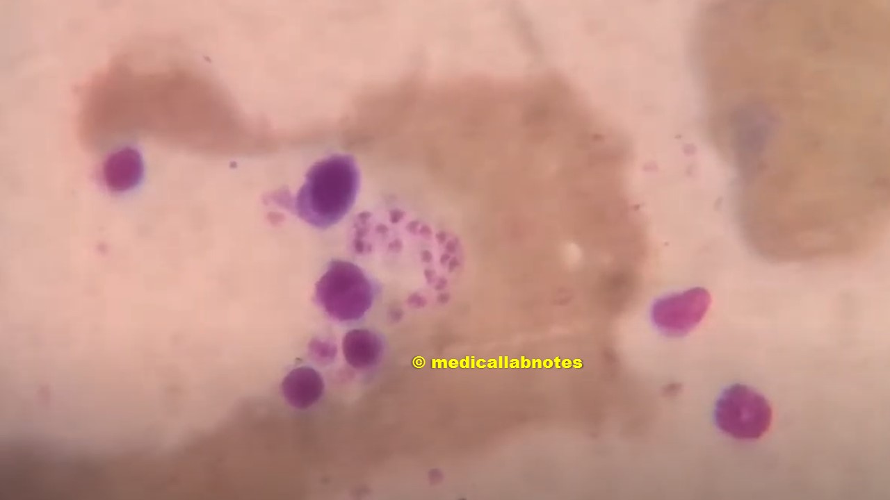
Introduction Pleural fluid is the liquid collected from the pleural space between the lung and chest wall. Microscopic examination of pleural fluid plays a key role in identifying the underlying cause of pleural effusion. It helps distinguish between transudates and exudates, detect infectious organisms, malignant …

Read more

Comparative Microscopy of Common Clinical Fungi-Introduction, Table, and Appearance at Different Magnifications

Aspergillus in LPCB tease mount of culture

Introduction Microscopy remains one of the cornerstones of clinical mycology, providing a rapid and inexpensive method for the direct detection and preliminary identification of fungi. Comparative microscopy is particularly valuable in distinguishing the morphological features of common clinical fungi, many of which exhibit overlapping clinical …

Read more

Diagnostic Challenge: LD Bodies vs Pneumocystis Vs Histoplasma in Giemsa-Stained BAL

Found in Giemsa staining of BAL-Possible pathogen?

Found in Giemsa staining of BAL. Possible pathogen? Morphological Features (Center) Possible Pathogen Supporting Features for Pneumocystis Recommendations Is it possible to be LD bodies? Based on the Giemsa-stained BAL image: The central violet-stained cluster of intracellular forms could resemble Leishman-Donovan (LD) bodies, especially in …

Read more

Trichomonas vaginalis: Introduction, Morphology, Pathogenicity, Lab Diagnosis, Treatment, Prevention, and Keynotes

Introduction Trichomonas vaginalis is a parasitic protozoan that primarily infects the urogenital tract of humans, causing the sexually transmitted infection (STI) known as trichomoniasis. This microorganism is responsible for one of the most common curable STIs worldwide. T. vaginalis was first identified by Alfred Francois …

Read more

Giemsa stain: Introduction, Principle, Reagent Preparation, Staining Procedure, Result Interpretation, Uses, and Keynotes

Leishmania donovani (LD bodies) amastigotes in Giemsa stained smear of bone marrow of a Visceral leishmaniasis (VL) or kala-azar patient

Introduction of Giemsa Stain Giemsa stain is a type Romanowsky stain that stains nuclei and cells. It was initially designed for the detection of malarial parasites in blood smears, but it is also used in histology for routine examination of blood smears. This technique uses for …

Read more