
Introduction of Giemsa Stain
Table of Contents
Giemsa stain is a type Romanowsky stain that stains nuclei and cells. It was initially designed for the detection of malarial parasites in blood smears, but it is also used in histology for routine examination of blood smears. This technique uses for the demonstration of other than malarial parasites, microbes like Helicobacter pylori, Chlamydia trachomatis, Borrelia species, Histoplasma capsulatum, Pneumocystis jiroveci, Penicillium marneffei and occasionally bacterial capsules and parasites like Toxoplasma gondii, Leishmiania donovani , Giardia lamblia, etc. It is also used to differentiate nuclear and cytoplasmic morphology of the various blood cells like RBCs, WBCs, and platelets. In cytogenetics, it stains the chromosomes and identifies chromosomal aberrations.
Principle of Giemsa Stain
Giemsa contains Methylene blue(AzureII)/Eosin. Methylene blue on oxidation produces colored compounds termed ‘Azure’ that have ability to combine with Eosin. Methylene blue azure is blue violet and stain acidic cell components while eosin is red and stains basic cell components.
Requirements
- Giemsa powder
- Glycerine
- Potassium dihydrogen
- phosphate
- Sodium hydroxide
- Methanol
- Weighing scale
- Measuring cylinder
- Conical flasks
- Glassware pipette
- Coplin jar
- Reagents bottles
- Gloves
- Masks
- Lab coat
- Cotton
Giemsa Stain Preparation
a. Preparation of Giemsa stain
| Ingredients | Volume/Amount |
| Giemsa stock powder | 0.5 gm |
| Glycerin | 27 ml |
| Methanol | 42 ml |
Giemsa powder is mixed in 27 ml of glycerin and pre-heated up to 600C. Then add methanol, shake the mixture and allow to stand for 7 days. Filter before use.
b) Buffer solution (stock)
| Ingredients | Amount/volume |
| Potassium dihydrogen phosphate | 1.36 gm |
| Distilled water | 50 ml |
| Sodium hydroxide | 0.4 gm |
| Distilled water | 50 ml |
- Dissolve both powder in distilled water.
- 50ml of potassium dihydrogen phosphate is mixed with 23.6ml of sodium hydroxide. The pH of the solution is adjusted to 6.8.
Working Solutions
Giemsa Stain: –
| Ingredients | Amount/Volume |
| Giemsa stock | 10 ml |
| Working buffer | 90 ml |
- The working Giemsa stain should be prepared fresh then use.
Buffer solution
| Ingredients | Amount/Volume |
| Stock buffer | 20 ml |
| Distilled water | 480 ml |
Staining Procedure
For Paraffin Section
- Deparaffinize and hydrated sections to tap water. Flood slide with Giemsa stain for 15-20 minute.
- Wash in tap water.
- Differentiate 0.2% acetic acid 1 dip.
- Wash in running tap water.
- Dehydrate, clear and mount with DPX.
For bone marrow imprints and smears
- Smears are fixed in methanol for 30 minutes.
- Smears are stained in working Giemsa solution for 20 minutes.
- Wash under running tap water for 5 minutes.
- Air dry smears, clear in xylene and mount in DPX.
Result-interpretation of Giemsa Staining
| Nucleus | Blue |
| Cytoplasm | Pink |
| H. pylori | Blue |
| LD bodies | Blue |
| Mast cell | Magenta pink |
| Tissue elements | Shades of blue to pink |
| Collagen, Muscles and Bone | Pale pink |
| Erythrocytes | Salmon pink |
| Malaria parasite | Malaria parasites have a red or pink nucleus and blue cytoplasm |
| Borrelia spirochetes | Mauve-purple |
| Chlymadia trachomatis inclusion bodies | Blue-mauve to dark purple depending on the stage of development |
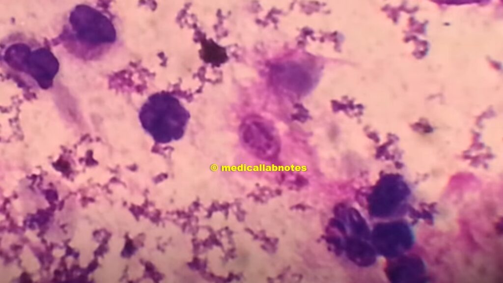
Leishmania donovani (LD bodies) amastigotes in Giemsa stained smear of bone marrow of a Visceral leishmaniasis (VL) or kala-azar patient

Cyst of Pneumocystis jiroveciin in Giemsa stained smear of BAL showing sporozoites
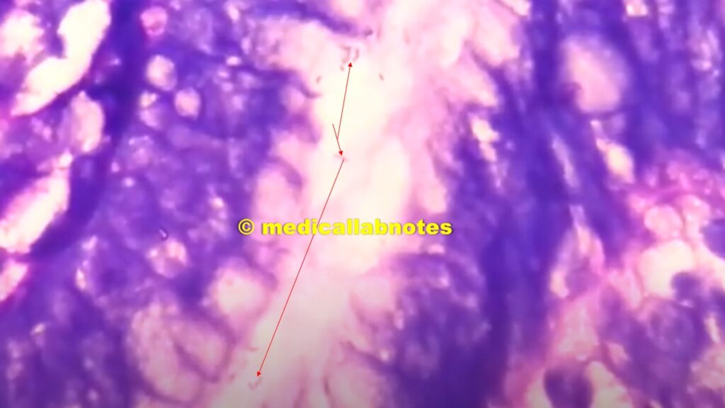
Helicobacter pylori in Giemsa stained gastric biopsy
Cryptococcus neoformans Capsules (clear zone covering body) in Giemsa stained smear of CSF

Uses of Giemas stain
- This stain is used for the demonstration of some microbes like Helicobacter pylori (bacteria) and parasites like Toxoplasma gondii, Leishmiania donovani , Giardia lamblia.
- It is also used to differentiate nuclear and cytoplasmic morphology of the various blood cells like RBCs, WBCs, and platelets.
- In cytogenetics, it stains the chromosomes and identifies chromosomal aberrations.
Keynotes
- Giemsa stain is the surname of German chemist and bacteriologist,Gustav Giemsa.
- Leishman stain differs from Jenner’s having eosin B instead of eosin Y.
- Occasionally bacterial capsules can also be noticed.
Bibliography
- Bailey and Scott’s Diagnostic Microbiology -13th Edn.
- Bancroft’s Theory and Practice of Histological Techniques (6th Edition)
- Mackie & Mc Cartney Practical Medical Microbiology- 14th Edn.
- Diagnostic Microbiology -Connie R. Mahon & George Manuselis
- Koneman Color Atlas and Textbook of Diagnostic Microbiology-6th Edn.
- Medical Microbiology-The Practice of Medical Microbiology Vol-2-12th Edn. –Robert Cruickshank
- District Laboratory Practice in Tropical Countries – Part-2- Monica Cheesebrough- 2nd Edn Update
I keep listening to the rumor lecture about getting free online grant applications so I have been looking around for the top site to get one. Could you tell me please, where could i acquire some?
Thank you for sharing with us, I think this website truly stands out : D.
Would love to constantly get updated outstanding blog! .
You have mentioned very interesting details! ps nice internet site. “‘We’re always lucky,’ I said and like a fool I did not knock on wood.” by Ernest Hemingway.
Hi there very nice site!! Guy .. Excellent .. Wonderful .. I’ll bookmark your site and take the feeds also…I am glad to find numerous useful info right here within the put up, we want work out more strategies on this regard, thanks for sharing. . . . . .
I’m no longer certain the place you are getting your information, however good topic. I needs to spend a while studying much more or understanding more. Thanks for wonderful info I was looking for this information for my mission.
I was very pleased to find this web-site.I wished to thanks for your time for this excellent learn!! I positively having fun with every little bit of it and I’ve you bookmarked to check out new stuff you weblog post.
certainly like your web-site but you have to check the spelling on several of your posts. A number of them are rife with spelling issues and I find it very bothersome to tell the truth nevertheless I will definitely come back again.
Thank you so much for providing individuals with an extraordinarily splendid opportunity to discover important secrets from this site. It can be very pleasant and also jam-packed with fun for me personally and my office mates to visit the blog nearly 3 times a week to study the fresh guidance you will have. And indeed, I’m usually astounded with your fantastic pointers you serve. Certain two ideas in this article are unquestionably the simplest we’ve ever had.
This really answered my problem, thanks!
Hi there! This post couldn’t be written any better! Reading through this post reminds me of my previous room mate! He always kept talking about this. I will forward this article to him. Pretty sure he will have a good read. Thank you for sharing!