
Introduction
Table of Contents
The name ‘ Atlas of Parasites’ is given even due to the vast spectrum of Parasitology but puny collection and another thing are that only an epic center collection of author authentical performance. So, please if you have benefited from this atlas, let others know about it too and share them through social media.

List of Contents
- Entamoeba histolytica/dispar in a saline wet mount
- Cyst of Entamoeba histolytica or dispar in an iodine wet mount
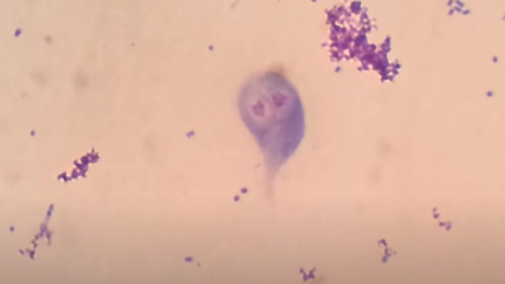
- Cyst and trophozoite of Entamoeba histolytica in LPCB wet mount
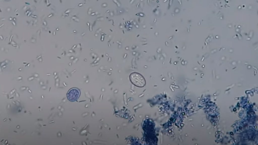
- Entamoeba coli cyst with 8 nuclei in iodine wet mount
- Giardia lamblia Cysts in Saline wet mount of Stool
- Cysts of Giardia lamblia in Iodine wet mount
- Methylene Blue wet mount of stool Microscopy showing cysts of Giardia lamblia
- Trichome stained smear of stool Microscopy showing Trophozoite of Giardia lamblia
- Trichomonas vaginalis
- Trichomonas hominis
- Blastocysts hominis
- Endolimax nana
- Cyclospora cayetanensis
- Cyclospora cayetanensis oocyst in modified Acid Fast staining
- Cryptosporidium parvum
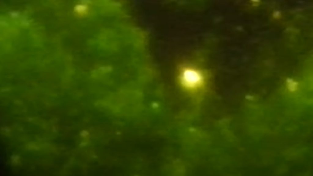
- Cryptosporidium parvum oocyst in Auramine phenol stain
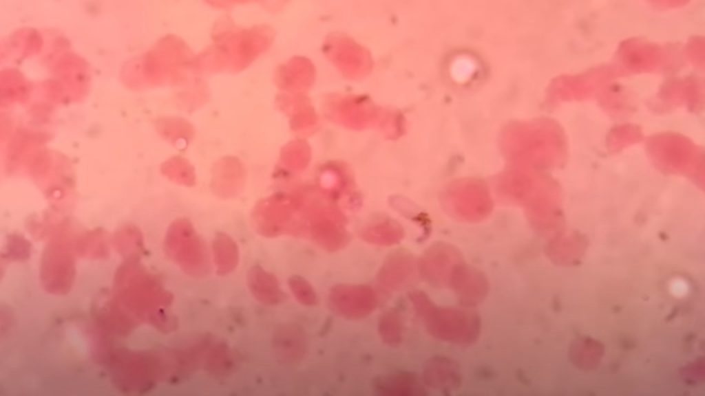
- Plasmodium species
- Leishmania donovani
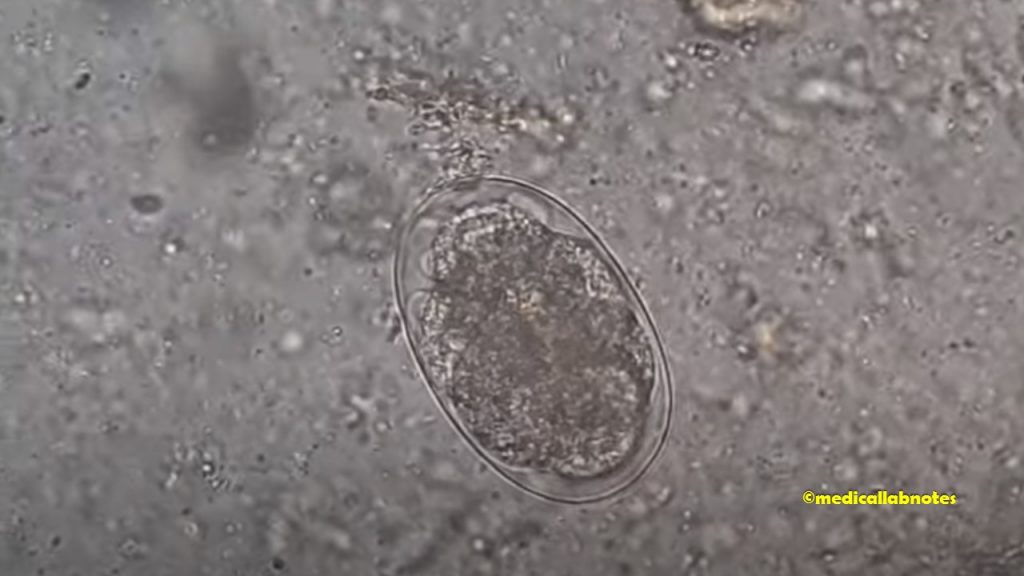
- Trichuris trichura or whipworm egg in a saline wet mount
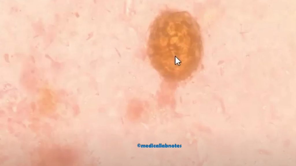
- Hookworm egg in an unstained wet mount
- Egg of tapeworm or Taenia in saline wet mount Microscopy
- Differentiating Taenia eggs found in human stool using Ziehl-Neelsen staining
- Ascaris or Roundworm egg in stool microscopy
- Fertilized egg changing to the larva of Ascaris or roundworm in stored stool microscopy
- Hymenolypsis nana or H. nana or Vampirolepsis egg showing hooklets in feces microscopy
- Hatching and Larval Release of Parasite
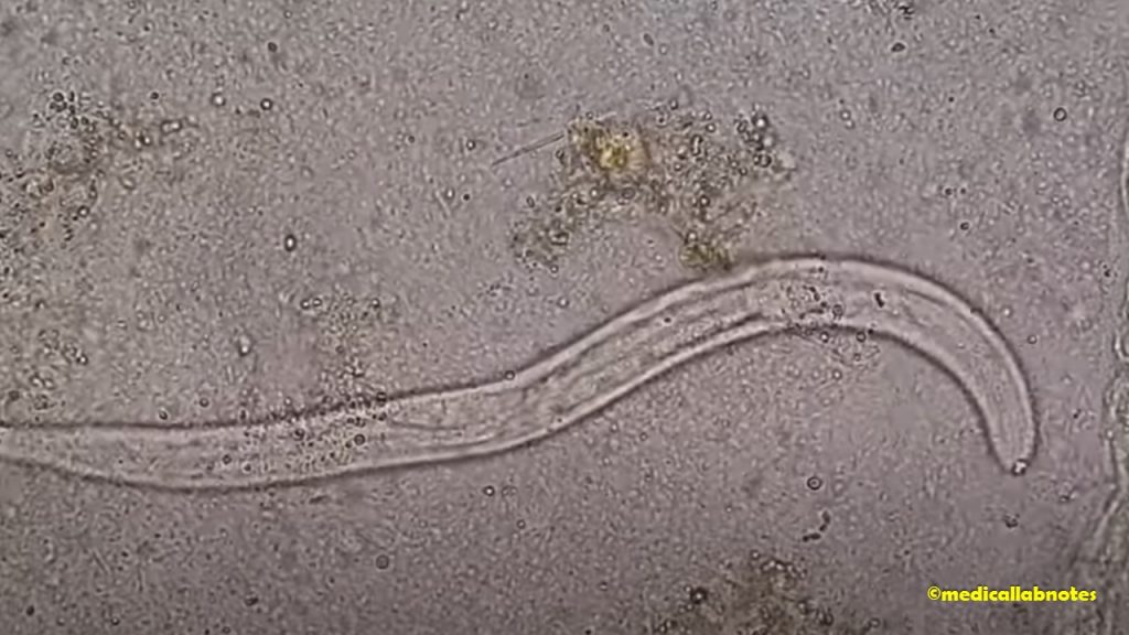
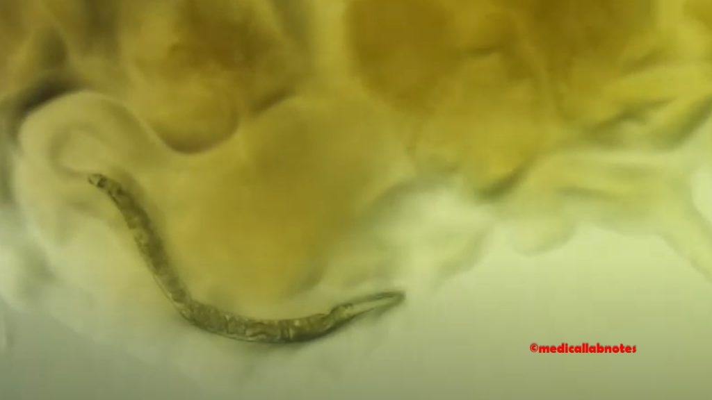
- Stool under the Microscope showing Parasite, Strongyloides stercoralis
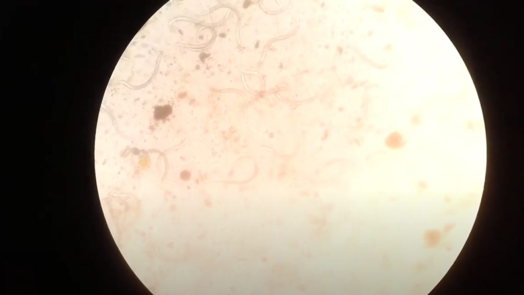
- Heavy load of parasites in the stool of a patient Microscopy
- Larva of the parasite, Strongyloides stercoralis in cultured MHA plate Microscopy
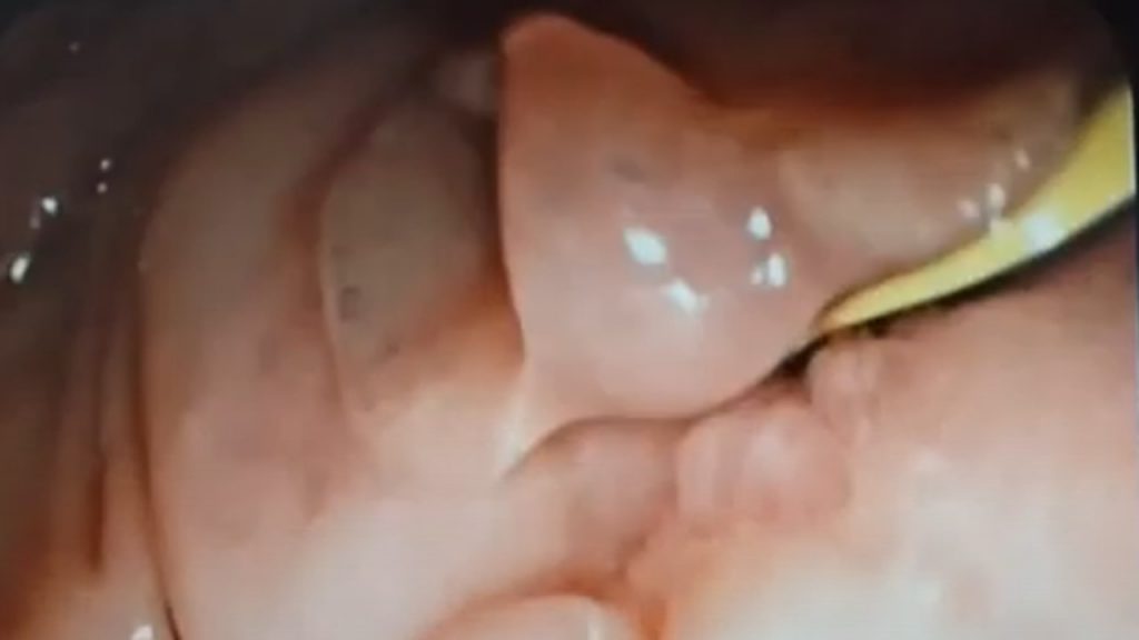
- Liver fluke or Fasciola encountered during ERCP -Liver fluke adult worm in the biliary passage of a patient
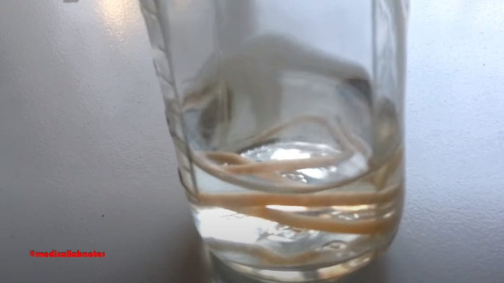
- Adult Ascaris lumricoides or roundworm demonstration
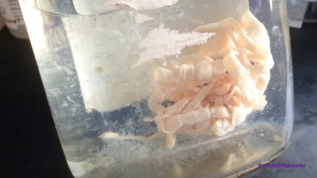
- Adult parasite-Tapeworm or Taenia Demonstration
- Dog Tapeworm, Dipylidium caninum
Description
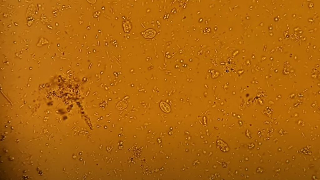
Entamoeba histolytica/dispar in a saline wet mount

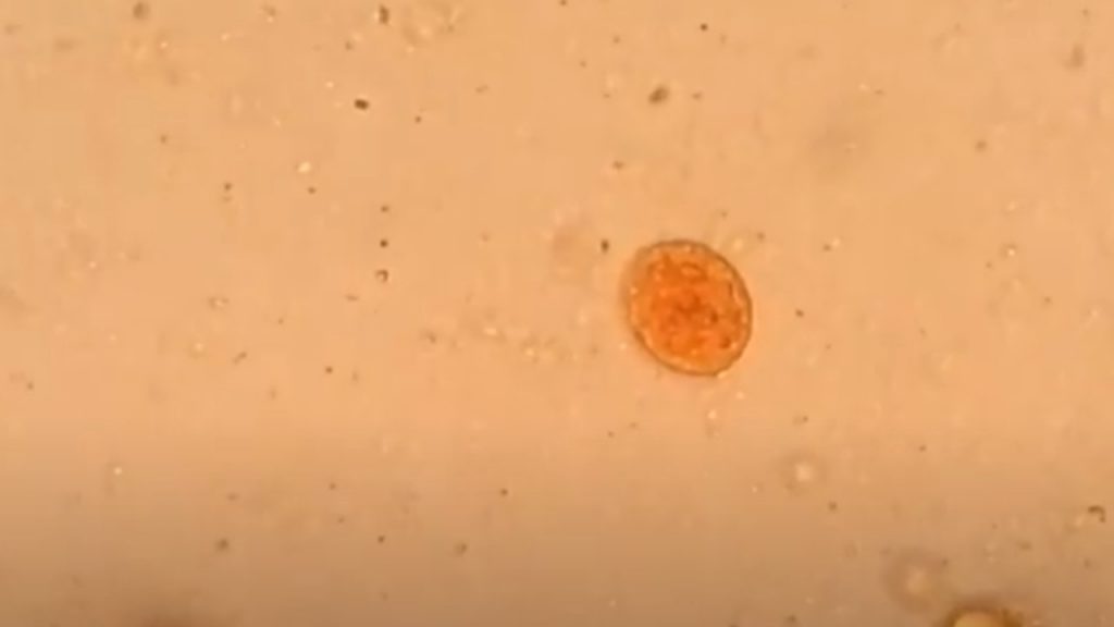
Cyst of Entamoeba histolytica or dispar in an iodine wet mount
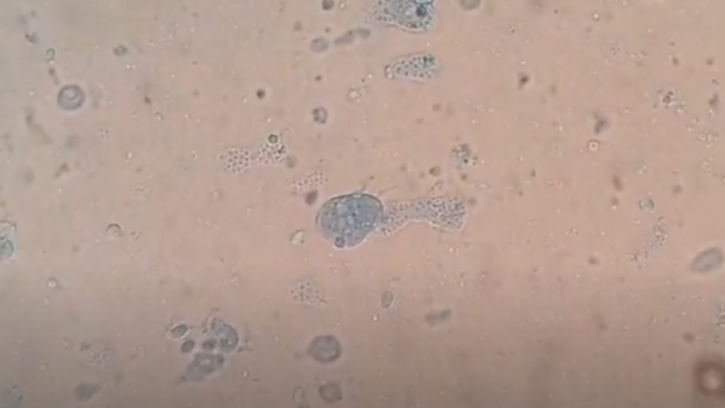
Cyst and trophozoite of Entamoeba histolytica in LPCB wet mount
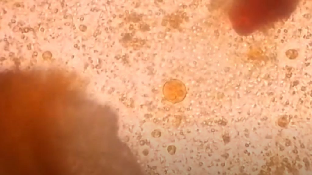
Entamoeba coli cyst with 8 nuclei in iodine wet mount
Giardia lamblia Cysts in Saline wet mount of Stool

Cysts of Giardia lamblia in Iodine wet mount
Methylene Blue wet mount of stool Microscopy showing cysts of Giardia lamblia
Trichrome stained smear of stool Microscopy showing Trophozoite of Giardia lamblia
Trichomonas vaginalis

Trichomonas hominis

Blastocysts hominis

Endolimax nana

Cyclospora cayetanensis

Cyclospora cayetanensis oocyst in modified Acid Fast staining

Cryptosporidium parvum

Cryptosporidium parvum oocyst in Auramine phenol stain
Plasmodium species
Leishmania donovani

Trichuris trichura or whip worm egg in saline wet mount

Hookworm egg in an unstained wet mount
Egg of tapeworm or Taenia in saline wet mount Microscopy

Differentiating Taenia eggs found in human stool using Ziehl-Neelsen staining
The Embryophore of this egg is lacking acid-fast and is also spherical rather than oval-shaped. So, we
can assume the egg may be of Taenia solium.

Ascaris or Roundworm egg in stool microscopy

Fertilized egg changing to the larva of Ascaris or roundworm in stored stool microscopy
Hymenolypsis nana or H. nana or Vampirolepsis egg showing hooklets in feces microscopy

Hatching and Larval Release of Parasite

Stool under the Microscope showing Parasite, Strongyloides stercoralis
Heavy load of parasites in the stool of a patient Microscopy
Larva of the parasite, Strongyloides stercoralis in cultured MHA plate Microscopy
Liver fluke or Fasciola encountered during ERCP -Liver fluke adult worm in the biliary passage of a patient
Adult Ascaris lumbricoides or roundworm demonstration
Adult parasite-Tapeworm or Taenia Demonstration
Dog Tapeworm, Dipylidium caninum
Many thanks to you for sharing all these wonderful threads. In addition, the right travel and also medical insurance system can often eradicate those issues that come with journeying abroad. The medical emergency can shortly become extremely expensive and that’s likely to quickly set a financial weight on the family finances. Setting up in place the excellent travel insurance offer prior to leaving is definitely worth the time and effort. Thanks a lot
I am continually searching online for articles that can assist me. Thank you!
Great – I should definitely pronounce, impressed with your website. I had no trouble navigating through all tabs as well as related info ended up being truly easy to do to access. I recently found what I hoped for before you know it at all. Reasonably unusual. Is likely to appreciate it for those who add forums or something, web site theme . a tones way for your customer to communicate. Excellent task..