Aspergillus niger Vs Aspergillus fumigatus-Introduction, Differences, Keynotes, and Related Footage

Aspergillus niger and Aspergillus fumigatus growth on Sabouraud dextrose agar (SDA) after 7 days of incubation

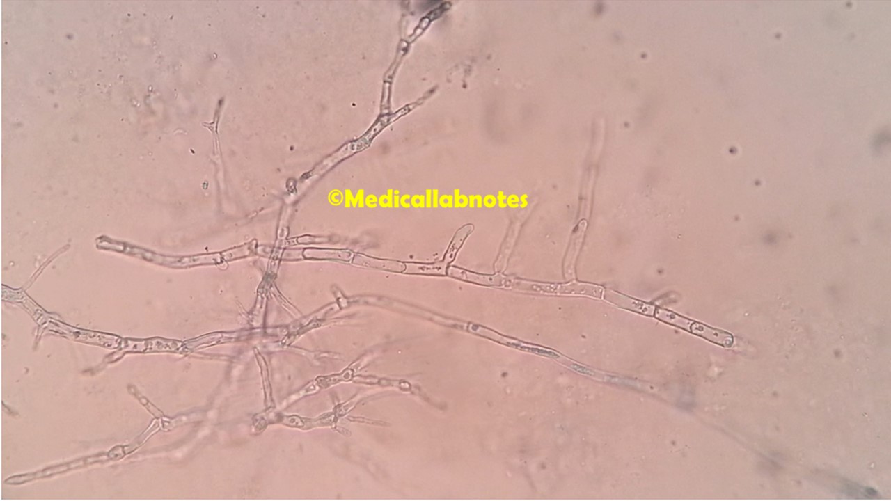
Introduction Key Differences: Aspergillus niger vs Aspergillus fumigatus Feature Aspergillus niger Aspergillus fumigatus Colony color (SDA) Black, powdery colonies Blue-green to gray-green colonies Growth rate Rapid, abundant sporulation Moderate, compact colonies Conidial heads Large, radiate, globose Columnar, compact Conidia Rough, black, spherical Smooth to rough, …

Read more

Comparative Microscopy of Common Clinical Fungi-Introduction, Table, and Appearance at Different Magnifications

Aspergillus in LPCB tease mount of culture

Introduction Microscopy remains one of the cornerstones of clinical mycology, providing a rapid and inexpensive method for the direct detection and preliminary identification of fungi. Comparative microscopy is particularly valuable in distinguishing the morphological features of common clinical fungi, many of which exhibit overlapping clinical …

Read more

Yeasts vs Molds: Detailed Comparison, Mixed Growth Isolation, Approach to Mixed Growth Handling, and Mixed Growth Interpretation

Yeast and mold mixed growth on SDA

Yeasts vs Molds: Detailed Comparison Feature Yeasts Molds Definition Unicellular fungi Multicellular filamentous fungi Structure Oval/round, single cells Hyphae form mycelium; septate or aseptate Reproduction Asexual (budding or fission); some sexual forms Asexual (spores like conidia/sporangia); sexual spores Hyphae Absent, but pseudohyphae may be present …

Read more

Bacteria and Fungi: Introduction, Description, Demonstration Techniques, Differences, and Keynotes

Bacteria and Fungi Introduction, Description, Demonstration Techniques, Differences, and Keynotes

Introduction of Bacteria and Fungi They are distinct types of microorganisms with different structures and characteristics. Bacteria are single-celled prokaryotes, while fungi can be single-celled (yeasts) or multicelled (molds, mushrooms) eukaryotes. They can be distinguished by their cell structure, colony morphology, and staining properties.  Description 1. Cell …

Read more

Fungal Microscopy: Introduction, List of Tests, Application, and Keynotes

Fungal Microscopy: Introduction, List of Tests, Application, and Keynotes

Introduction Fungal microscopy visualizes fungi in clinical specimens. Moreover, it diagnoses fungal infections rapidly.Additionally, it identifies fungal morphology and structures. Furthermore, it guides antifungal therapy decisions. Consequently, it plays a crucial role in clinical mycology. List of Tests for Fungal Microscopic Examination Firstly, the KOH …

Read more

Slide Culture Technique for Fungi- Introduction, Requirement, Procedure, Uses, Disadvantage, and Keynotes

Slide Culture Technique for Fungi- Introduction, Requirement, Procedure, Uses, Disadvantage, and Keynotes

Introduction The Slide Culture Technique for fungi allows detailed observation of fungal structures. It provides a clear view of spores and hyphae. This method uses a small block of agar on a slide. The technique is beneficial for identifying molds. You place a coverslip on …

Read more

Microsporum ferrugineum: Introduction, Morphology, Pathogenicity, Lab Diagnosis, Treatment, Prevention, and Keynotes

Microsporum ferrugineum in LPCB taese mount of culture microscopy showing typical bamboo hyphae and lacking production of both microconidia and macroconidia

Introduction Microsporum ferrugineum is a species of dermatophytic fungus that belongs to the genus Microsporum. Dermatophytes are a group of fungi that are capable of infecting and colonizing the outer layers of the skin, hair, and nails, causing various dermatophytosis or “ringworm” infections in humans …

Read more