
Introduction of Germ Tube
Table of Contents
The Germ Tube Test procedure is used for the presumptive identification of Candida species. The culture of Candida species is treated with normal human pooled serum or sheep or rabbit or fetal calf serum and incubated at 37°C for 2-4 hours. A drop of suspension is examined on the slide under a microscope. The germ tubes are seen as long as tube-like projections extending from the yeast cells. There is no constriction at the point of attachment to the yeast cell as seen in the case of Pseudohypahe. The demonstration of the germ tube is known as the Reynolds-Braude phenomenon. This is a rapid method for identifying and differentiating C. albicans from other Candida spp. Buds and pseudohyphae can be distinguished from germ tubes by the constricted attachment at the point of origin. Germ tubes don’t show constriction at the point of origin.
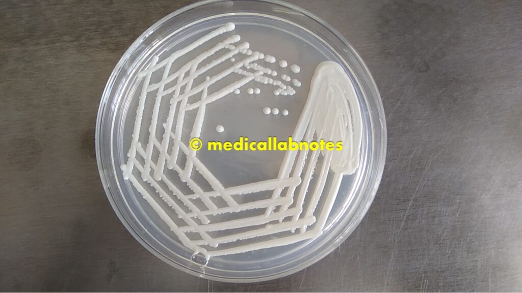
Candida albicans growth on SDA
Candida albicans growth on Chromagar

Principle of Germ Tube Test
A germ tube is a short outgrowth, non-septate germinating hypha. It is ½ the width and 3 – 4 times the length of the cell from which it arises. When Candida species are incubated in serum at 37°C for 2-4 hours and produce short, slender, tube-like structures called germ tubes. The formation of this germ tube is associated with increased synthesis of protein and ribonucleic acid and is observed in Candida albicans.
Requirements for Germ Tube Test
- Test tube (12Χ75 mm)
- Inoculating loop/ sterile bamboo stick
- 24 hours old culture of the suspected fungal colony to be tested
- Incubator
- Clean and grease-free slide and coverslip
- droppers
- Microscope
- Human pooled serum
- Control strains-Positive control (PC): Candida albicans while Negative control (NC): Candida parasilosis
Procedure of Germ Tube Test
- Take four test tubes and label them as uninoculated (UN), negative control (NC), positive control (PC), and test (T)
- Add 0.5 ml of serum to each test tube.
- Take half of a single colony to be tested by using a sterile loop, and mix with serum in the test tube ‘T’.
- Similarly, take half of C. albicans single colony by using a sterile loop, and mix it with serum in the test tube PC.
- Similarly, take half of C. parasilosis single colony by using a sterile loop, and mix it with serum in the test tube NC.
- Leave an uninoculated tube (UN) without any disturbance.
- Now incubate all the tubes at 37°C for 2 hours.
- Place one drop of suspension from tube tubes Un, NC, PC, and T onto different slides and place cover slips over the drops.
- Examine the slide under low power (10X) and finally high power (40X) objectives.
Observations of Germ Tube Test
- Under the microscope, the whole field of the coverslip is examined for any yeast cell showing the production of a germ tube.
- Germ tubes are seen as long tube-like projections extending from yeast cells and this should be differentiated from pseudohyphae.
- Differences between germ tube and pseudohyphae-
| Germ Tube | Pseudohyphae |
| No constriction at the site of attachment. | Constriction at the site of the attachment |
| Non-septate with parallel sides. | Septate and not necessarily with parallel sides. |
Result Interpretation of Germ Tube Test

- Positive Result: A short hyphal (filamentous) extension arising laterally from a yeast cell with no constriction at the point of origin. May be confirmed as C. albicans
- Negative Result: No hyphal extension arising from a yeast cell or a short hyphal extension with constriction at the point of origin.
- Uninoculated (UN) tube: Lacking yeast cells
- Negative Control (NC) tube: Absence of germ tube formation
- Positive Control(PC): Presence of germ tubes as shown above figure.
- The exact mechanism of Germ tube formation is still unknown.
Keynotes on Germ Tube Test
- A germ tube is a useful test to identify C. albicans and a few other species which are pathogenic.
- Candida tropicalis may produce pseudo-germ tubes after 3 hrs of incubation but they show constriction at the point of origin.
- Rabbit, fetal calf, or human serum can be used for demonstration of germ tube formation.
- Too heavy inoculum will inhibit germ tube formation.
- Germ tubes are produced by C. albicans and C. dubliensis and one test for distinguishing C. dubliniensis from C. albicans, is a laboratory culture of the organism at 42 °C. Most C. albicans strains grow at this temperature, whereas most C. dubliniensis isolates do not.
- At times, some strains of C. albicans isolated from patients with antifungal drugs or patients with cancer do not produce germ tubes.
Candida albicans Colony Morphology on SDA

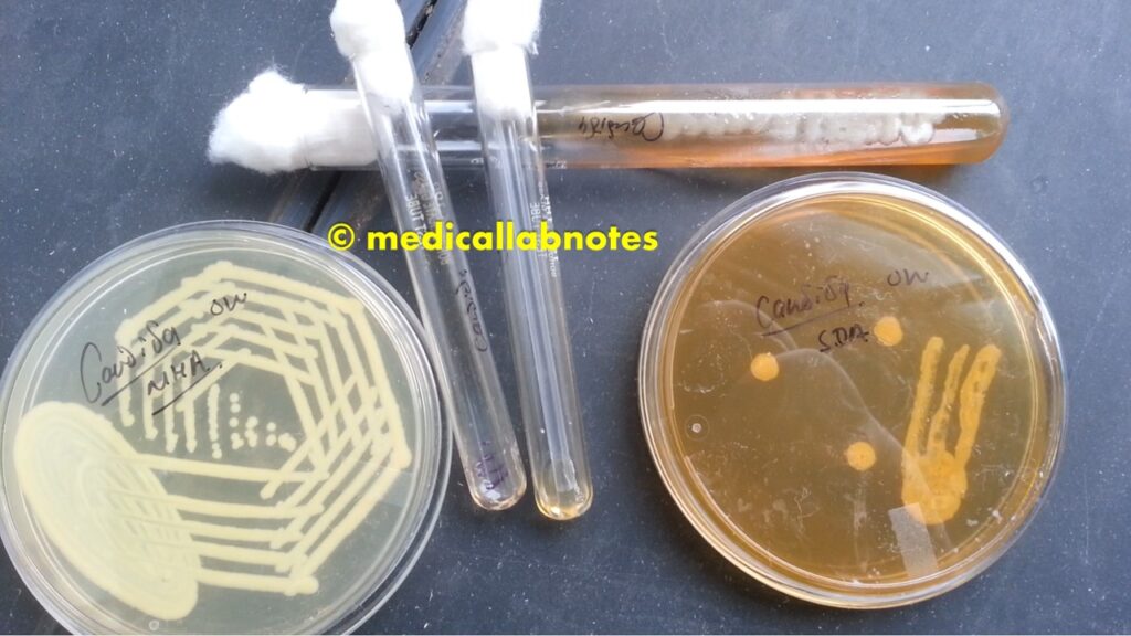
Candida species on various culture media (MHA, SDA, Chromagar)
Candida albicans under a Fluorescence microscope stained with Acridine Orange

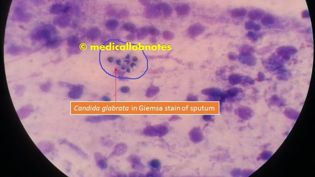
Candida glabrata in Giemsa stained smear of sputum
Yeast cells and Pseudohyphae in Gram Staining of Sputum

Further Readings
- A textbook of Medical Mycology-Jagdish Chander
- Description of Medical Fungi-David Ellis, Stephen Davis, Helen Alexiou, Rosemary Handke, Robyn Bartley
- Textbook of Practical Microbiology-Subhash Chandra Parija