All Notes

Fungal Elements in KOH Mount of BAL (Bronchoalveolar Lavage) Microscopy- Introduction, Fungal Elements Observed in BAL KOH Mount, Applications, and Keynotes

Introduction

KOH mount (Potassium Hydroxide preparation) is a rapid, simple, and cost-effective microscopic technique used to detect fungal elements directly in clinical specimens.
When applied to Bronchoalveolar Lavage (BAL) samples, it is an essential diagnostic tool for identifying pulmonary fungal infections, especially in the ICU, oncology, transplant, and immunocompromised patients.

Fig. BAL (Bronchoalveolar Lavage) for KOH mount Microscopy-

KOH dissolves mucus, cellular debris, and proteins present in the BAL fluid, clearing the background while

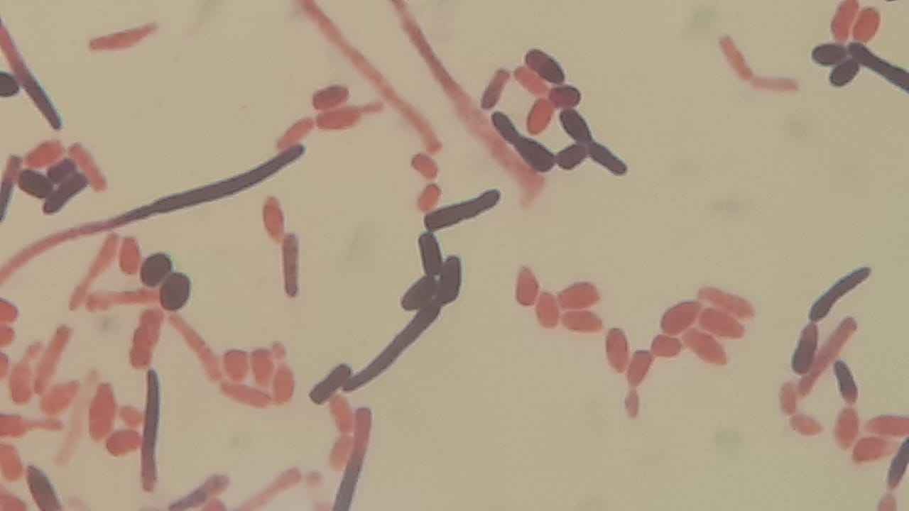
Fig. Fungal Elements in KOH Mount of BAL (Bronchoalveolar Lavage) Microscopy at 40X objective

, preserving the chitin-rich fungal cell walls, making fungal structures easily visible under the microscope.

Fig. Fungal Elements in KOH Mount of BAL (Bronchoalveolar Lavage) Microscopy

BAL KOH mount is extremely helpful in diagnosing invasive fungal infections, opportunistic mycoses, and mixed fungal–bacterial infections in the lungs.

Fungal Elements Observed in BAL KOH Mount

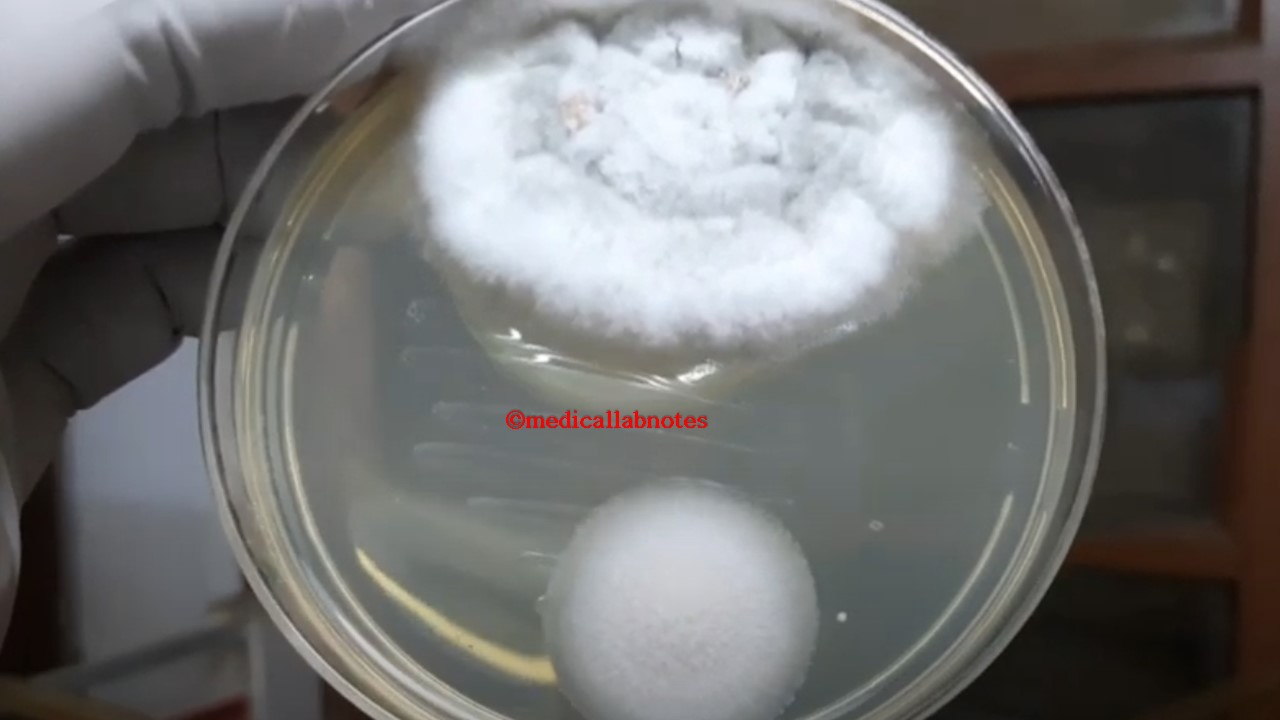
Fog. BAL (Bronchoalveolar Lavage) for KOH mount and fungal culture

1. Yeast Cells

  • Round/oval, refractile structures
  • May show budding (blastoconidia)
  • Common species: Candida, Cryptococcus
  • Cryptococcus may show a halo-like capsule clearing

2. Pseudohyphae

  • Elongated, sausage-like, with constrictions at septa
  • Seen commonly in Candida albicans
  • Indicates active infection rather than colonization

3. True Hyphae

4. Conidia / Spores

5. Pneumocystis jirovecii Structures (Occasional)

  • Foamy alveolar casts
  • Crushed “ping-pong ball” cysts
  • Better visualized with GMS or Giemsa, but KOH may show cyst-like bodies

Applications of Fungal Elements in KOH Mount of BAL (Bronchoalveolar Lavage) Microscopy

  • Rapid Screening Test: Provides immediate detection of fungal elements before culture or molecular tests.
Fig. Fungal growth on Sabouraud dextrose agar after 2 days of incubation at 37 °C
  • Diagnosis of Pulmonary Fungal Infections: Useful in identifying invasive aspergillosis, mucormycosis, pneumocystis pneumonia, and Candida pneumonia.
  • Critical Care and Immunocompromised Patients: Essential for diagnosing fungal infections in cancer, HIV/AIDS, transplant, and ICU patients.
  • Guiding Antifungal Therapy: Helps clinicians initiate early antifungal treatment, especially in emergency settings.
  • Corroborating Culture and Molecular Findings: Microscopy supports results from culture, PCR, sequencing, or antigen tests (e.g., galactomannan, β-D-glucan).

Keynotes on Fungal Elements in KOH Mount of BAL

Fig. Fungal Elements in the wet mount of the culture of a BAL (Bronchoalveolar Lavage) specimen
  1. KOH concentration used: 10–20%
  2. Fungal elements appear refractile, clear, and well-defined against a cleared background
  3. BAL KOH mount is rapid but not species-specific—confirm with culture or molecular tests
  4. Helpful for differentiating colonization versus invasive infection when correlated clinically
  5. Essential for diagnosing fungal pneumonia in oncology, ICU, and immunocompromised patients
  6. Should be interpreted by an experienced microbiologist to avoid artifacts (e.g., mucus threads, crystals)
  7. Always pair with SDA culture, GMS stain, PAS stain, or PCR for confirmation
Fig. Fungal Elements in the wet mount of the culture of the BAL specimen
Fig. Fungal elements in the wet mount of the culture of BAL fluid

Further Readings

  1. https://pmc.ncbi.nlm.nih.gov/articles/PMC10492593/
  2. https://www.researchgate.net/figure/KOH-mount-of-bronchoalveolar-lavage-fluid-obtained-after-bronchoscopy-Magnification-3440_fig3_23655567
  3. https://pmc.ncbi.nlm.nih.gov/articles/PMC10492593/#:~:text=On%20the%20other%20hand%2C%20many,to%20provide%20more%20appropriate%20treatment.
  4. https://www.researchgate.net/figure/KOH-mount-of-bronchoalveolar-lavage-fluid-obtained-after-bronchoscopy-Magnification-3440_fig3_23655567
  5. https://www.1mg.com/labs/test/koh-staining-sputum-35070
  6. https://www.dalynn.com/dyn/ck_assets/files/tech/RP85-25.pdf
  7. https://drkothiwalaskineva.com/medical-dermatology/koh-mount-for-fungal-diseases/
  8. https://ijdvl.com/skin-scraping-and-a-potassium-hydroxide-mount/
  9. https://ritm.gov.ph/laboratory-services/fungal-culture-and-susceptibility-test-urineinclusion1-potassium-hydroxide-mount-koh-29/
  10. https://en.wikipedia.org/wiki/KOH_test#:~:text=Collection:%20Skin%2C%20nail%2C%20or,follicles%20from%20the%20infected%20site.
  11. https://www.ncbi.nlm.nih.gov/books/NBK430762/

Medical Lab Notes

Recent Posts

Orientia and Rickettsia: Introduction, Comparison, Key Differences, Treatment and Diagnosis

Introduction Orientia and Rickettsia are two closely related genera of bacteria within the family Rickettsiaceae.…

11 hours ago

Biosafety Officer: Introduction, Roles, Uses, Clinical Significance, and Keynotes

Introduction A Biosafety Officer (BSO) is a technical expert responsible for the strategic and integrated…

13 hours ago

Immunohistochemistry (IHC): Introduction, Application, and Keynotes

Introduction to IHC Immunohistochemistry (IHC) is a powerful laboratory technique that combines anatomical, immunological, and…

1 day ago

Tru-Cut Biopsy (Core Needle Biopsy): Introduction, Procedure, Application, and Keynotes

Introduction A Tru-cut biopsy, also known as a core needle biopsy (CNB), is a minimally…

1 day ago

VITEK 2 Yeast AST System: Introduction, Name of Card, Principle, Procedure, Result-Interpretation, and Keynotes

Introducton The VITEK 2 Yeast AST system is a fully automated solution for the quantitative…

1 day ago

VITEK 2 Yeast Identification: Introduction, Name of Card, Principle, Procedure, Result-Interpretation, Organisms Identified, and Keynotes

Introduction The VITEK 2 Yeast Identification (YST) system is an automated platform designed for the…

1 day ago