Introduction
Table of Contents
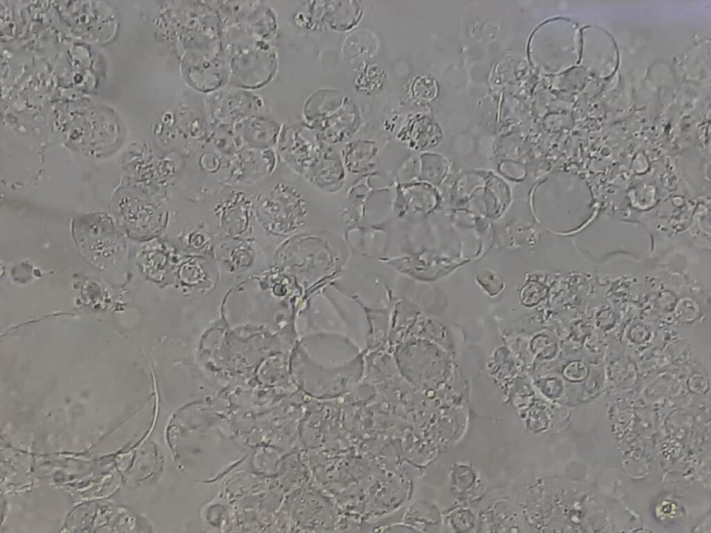
Microscopic evaluation of ascitic fluid is an essential step in differentiating between benign lipid material and pathological cellular elements. The distinction between fat globules and cells is critical because both may appear as rounded structures, yet their origin, clinical significance, and interpretation differ significantly.
Fat Globules in ascitic fluid usually arise from lipid leakage following trauma, pancreatitis, chylous ascites, or malignancy-induced lymphatic obstruction. Under the microscope, they appear as clear, refractile, variably sized droplets that may float freely in the background without internal structures. They lack nuclei and do not stain with routine cytological dyes, but may demonstrate special staining with fat-specific stains like Sudan III or Oil Red O.
Cells, on the other hand, represent cellular components of ascitic fluid such as mesothelial cells, inflammatory cells (neutrophils, lymphocytes, macrophages), or malignant cells. Unlike fat globules, cells exhibit definite nuclear and cytoplasmic details, take up stains like Giemsa or Papanicolaou, and often demonstrate arrangement patterns (clusters, sheets, or single forms).
Comparison
| Feature | Fat Globules (Lipid Droplets) | Cells (Mesothelial/Inflammatory/Malignant) |
| Size | Variable, often large (up to 100 µm) | Smaller, more uniform (5–20 µm) |
| Shape | Round, refractile, clear borders | Round/oval; may be irregular in malignant cells |
| Refractility | Strongly refractile, shiny | Less refractile, dull appearance |
| Nucleus | Absent | Present (single, multiple, or atypical nuclei) |
| Cytoplasm | May fuse, form larger droplets | Granular or vacuolated, sometimes basophilic |
| Clustering | Float and move when the slide is tilted | Seen in clusters (mesothelial cells) or groups (malignant cells) |
| Staining | Poorly stained with routine stains | Take up stains (e.g., Giemsa, Pap, H&E) |
| Mobility (wet prep) | Float and move when slide is tilted | Stationary, firmly settled |
| Clinical Significance | Suggests chylous/pseudochylous ascites (lymphatic obstruction, TB, lymphoma, trauma) | Indicates inflammation, infection, or malignancy |
| Special Tests | Sudan III/IV, Oil Red O → positive for fat | Cytology, culture, immunocytochemistry → to characterize cell type |
Keynote
- In ascitic fluid microscopy, the presence of fat globules points toward chylous ascites, while cells (mesothelial, neutrophils, lymphocytes, or malignant) give clues to infection, TB, or cancer. Correct differentiation is essential for diagnosis.