
Introduction
Table of Contents
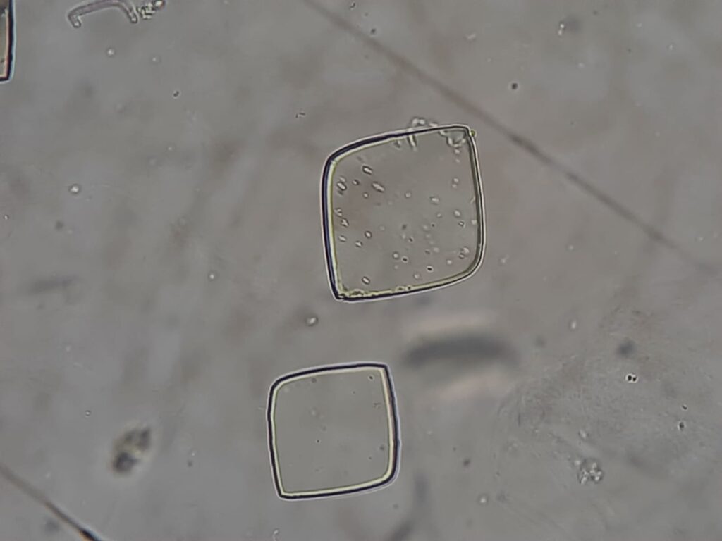
Urate crystals, also referred to as uric acid crystals, are metabolic by-products of purine metabolism found in urine. They are frequently observed during urine microscopy, especially in patients with altered pH, dehydration, or metabolic disorders. Their presence can be physiological in concentrated urine or pathological in cases linked to hyperuricemia, gout, or renal dysfunction.


Identification Features

- Shape: Typically rhomboid, diamond, or rosette-shaped.
- Edges: Sharp and well-defined.
- Color (under light microscopy): Often yellow to reddish-brown, but may appear colorless.
- Polarization: Exhibit birefringence under polarized light.
- Urine pH: Commonly found in acidic urine (pH < 6.0).
- Magnification: Clear visualization at 100X and 400X magnification.
- Differentiation: Must be distinguished from calcium oxalate crystals (which appear as envelope/dumbbell-shaped and are found in acidic to neutral urine).

Clinical Significance
- Normal finding:
- It may be seen in healthy individuals due to dehydration, high-protein diets, or fasting.
- Pathological associations:
- Gout: Excess uric acid deposition leading to hyperuricemia.
- Renal calculi (uric acid stones): Predisposed by low urine pH and high urate concentration.
- Tumor lysis syndrome: The rapid breakdown of tumor cells releases purines, leading to an increase in uric acid.
- Chemotherapy/radiotherapy: Similar mechanism to tumor lysis syndrome.
- Chronic kidney disease: Impaired uric acid excretion.
- Diagnostic importance:
- Serves as a microscopic clue in metabolic workups for disorders of uric acid metabolism.
- Helps distinguish urate crystals from infectious or other metabolic crystals.

Keynotes
- Urate crystals indicate acidic urine and may or may not suggest pathology.
- Differentiation from calcium oxalate and cystine crystals is vital for correct interpretation.
- Clinicians should correlate microscopy findings with serum uric acid levels, renal function tests, and clinical symptoms.
- Persistent presence warrants evaluation for gout or renal stone formation.
Further Readings
- https://www.ncbi.nlm.nih.gov/books/NBK606103/
- https://www.sciencedirect.com/topics/medicine-and-dentistry/urate-crystal
- https://eclinpath.com/urinalysis/crystals/
- https://dreminozbek.com/en/crystaluria-causes-types-and-treatment/
- https://medlineplus.gov/lab-tests/crystals-in-urine/
- https://en.wikipedia.org/wiki/Uric_acid
- https://onlinelibrary.wiley.com/doi/10.1002/jcla.24707
- https://emedicine.medscape.com/article/983759-overview
- https://www.frontiersin.org/journals/medicine/articles/10.3389/fmed.2021.649505/full