The name ‘ Atlas of Bacteria’ is given even due to the vast spectrum of bacteriology but puny collection and another thing is that only an epic center collection of author authentical performance. So, please if you have benefited from this atlas, let others know about it too and share them through social media.
Atlas of Bacteria: Introduction, List of Contents, and Description
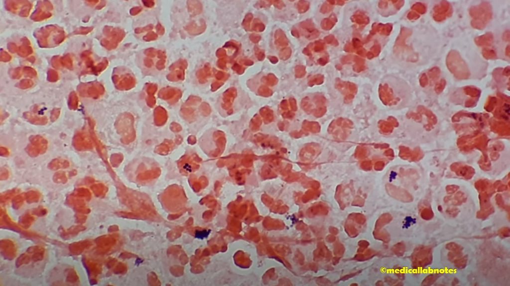
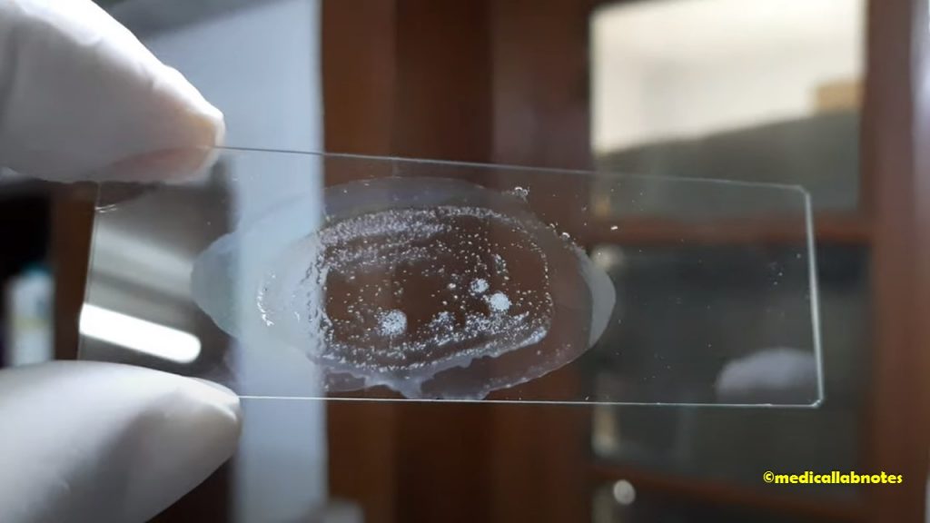
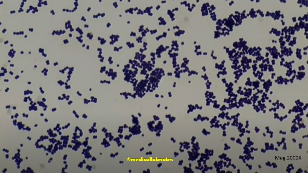
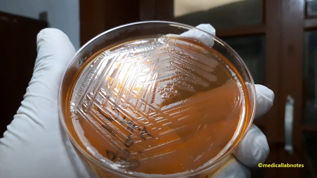
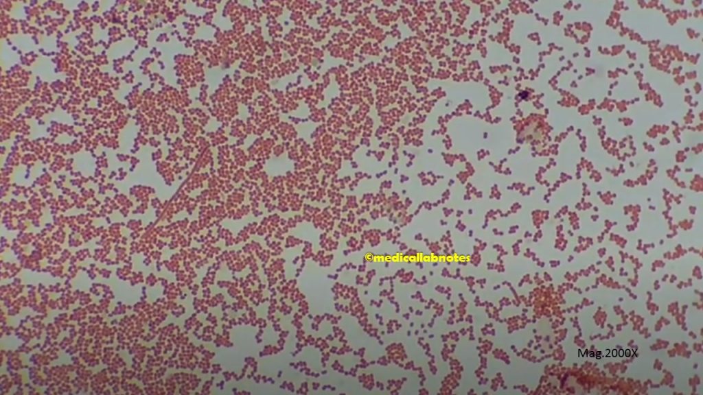
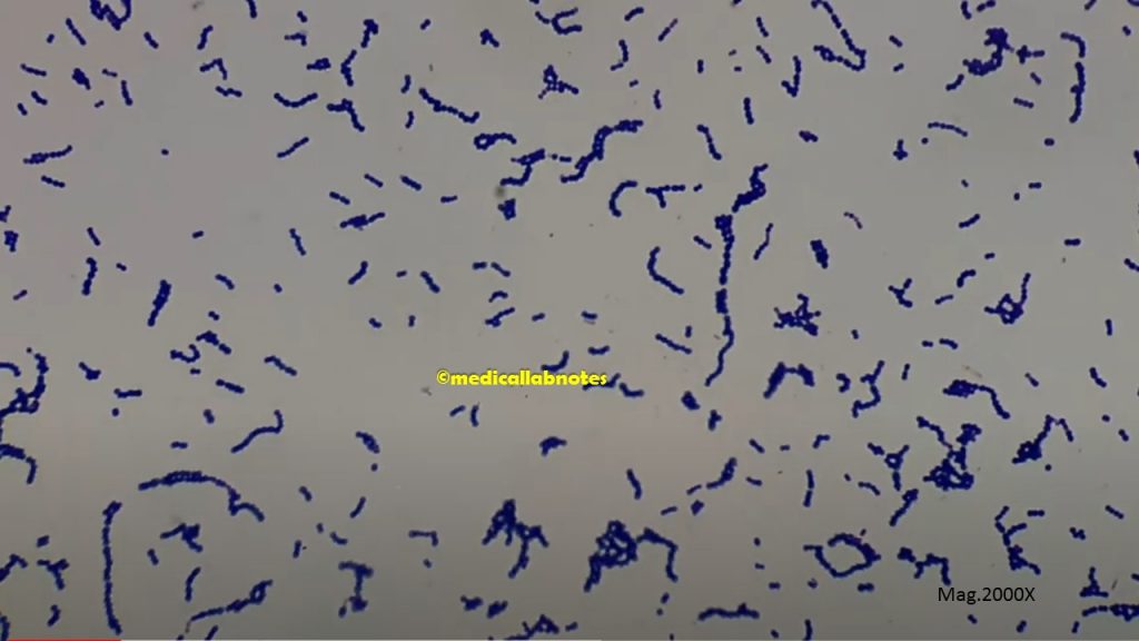
Aeromonas species in Gram-stained of culture Microscopy
Fig. Aeromonas species in Gram-stained of culture Microscopy
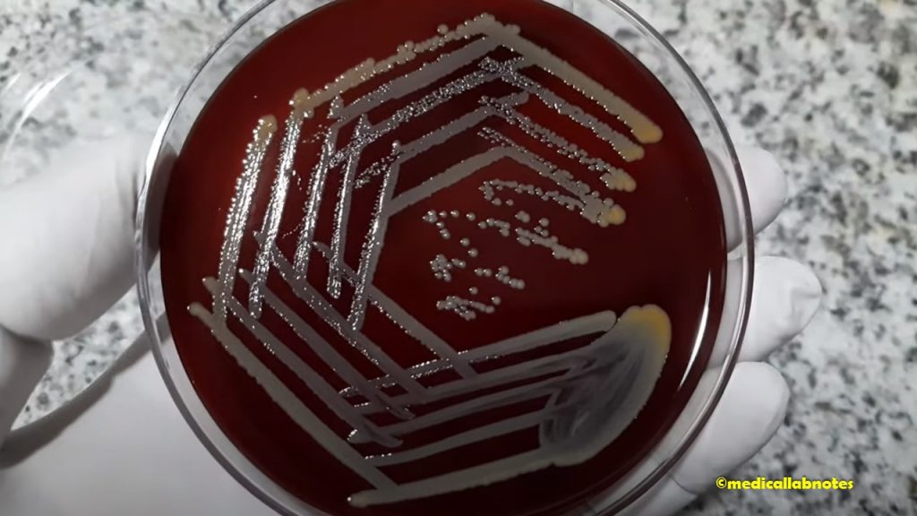
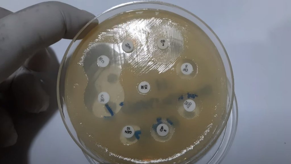
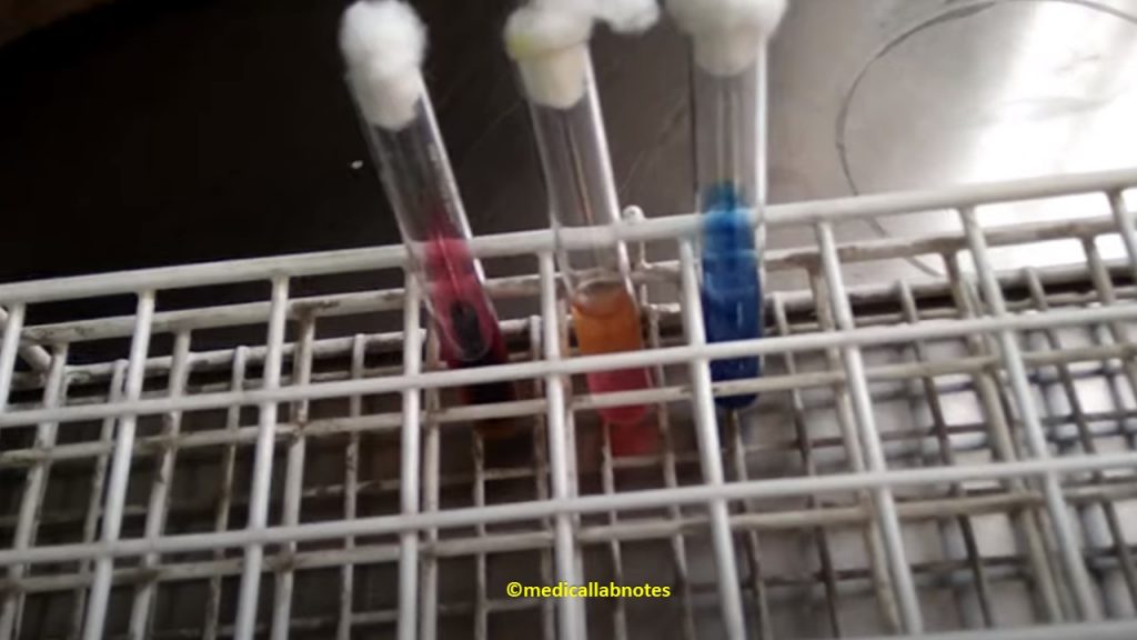
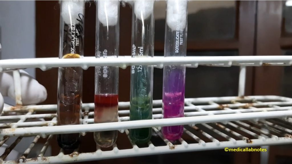
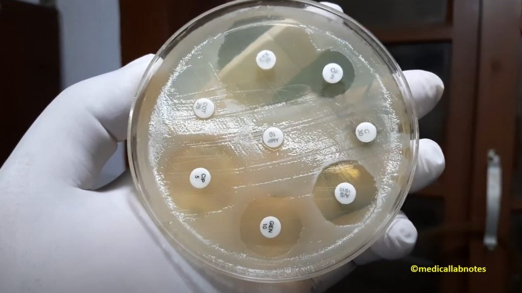
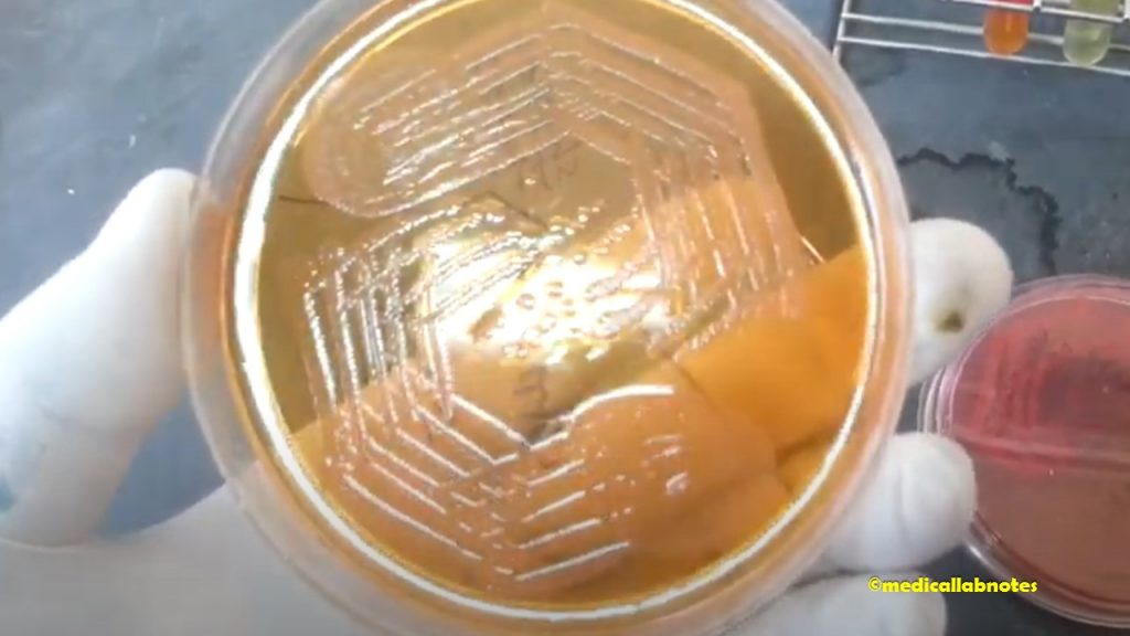
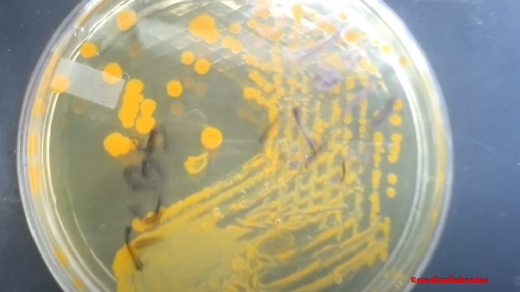
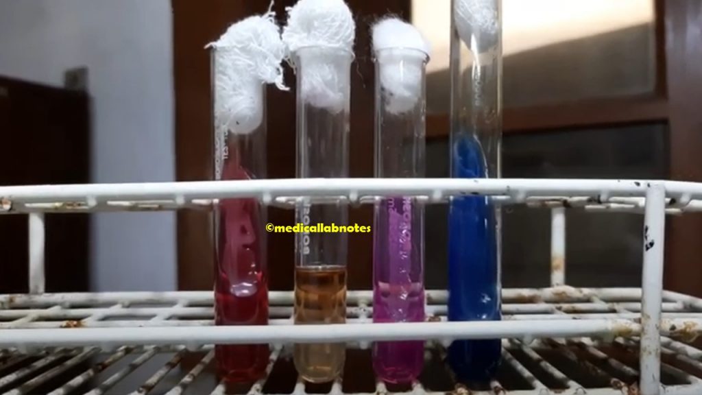
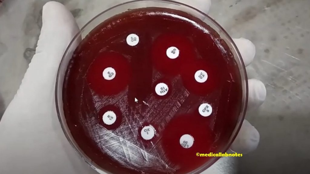
Aeromonas species Antibiogram
Fig. Aeromonas species Antibiogram
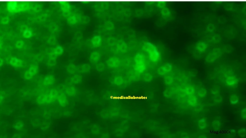
Haemophilus influenzae in Gram-stained smear of Pneumonia patient sputum Microscopic Footage
Fig. Haemophilus influenzae in Gram-stained smear of Pneumonia patient sputum Microscopic Footage at a magnification of 1000X showing Gram-negative coccobacilli, small to large rods with a background of pus cells
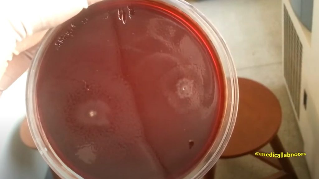
Use of Bacitracin (10 Units) disk in blood agar for screening Haemophilus species
Fig. Use of Bacitracin (10 Units) disk in blood agar for screening Haemophilus species
Haemophilus influenzae growth on chocolate agar
Fig. Haemophilus influenzae growth on chocolate agar
Haemophilus influenzae in Gram-stained smear of culture showing Gram-negative coccobacilli, small to large rods
Fig. Haemophilus influenzae in Gram-stained smear of culture showing Gram-negative coccobacilli, small to large rods
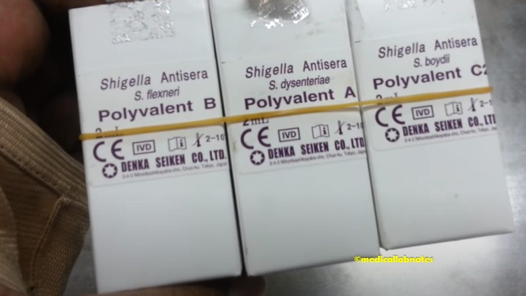
Haemophilus influenzae Antisera
Fig. Haemophilus influenzae Antisera
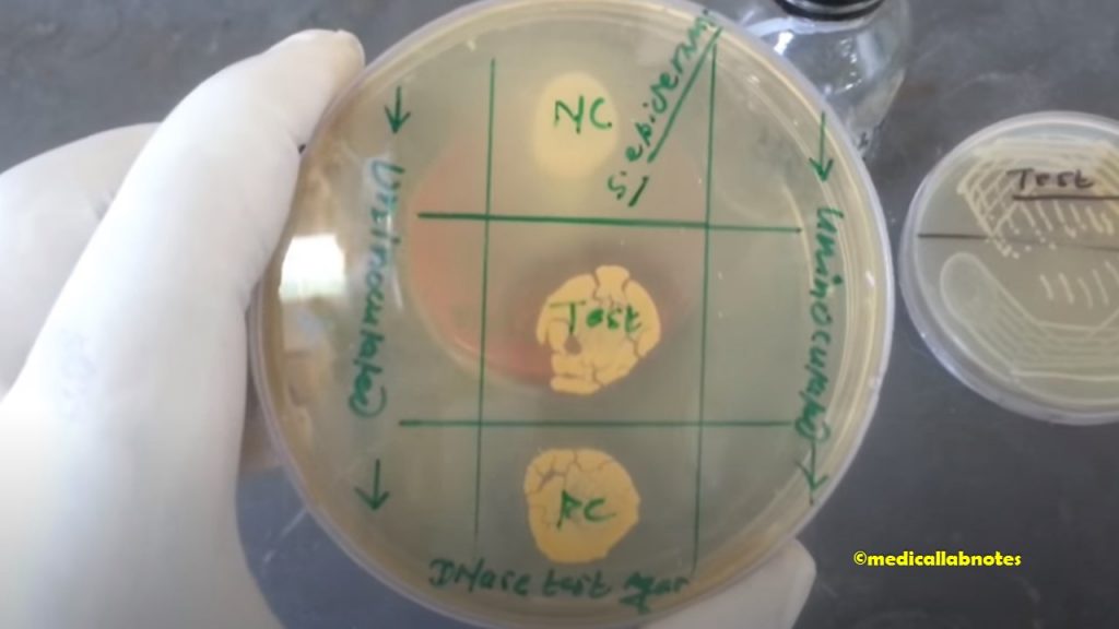
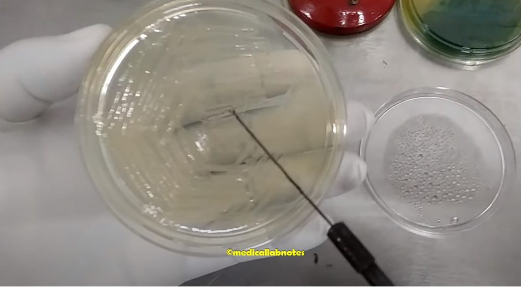
Satellitism Test for the Identification of Haemophilus influenzae showing positive
Fig. Satellitism Test for the Identification of Haemophilus influenzae showing positive
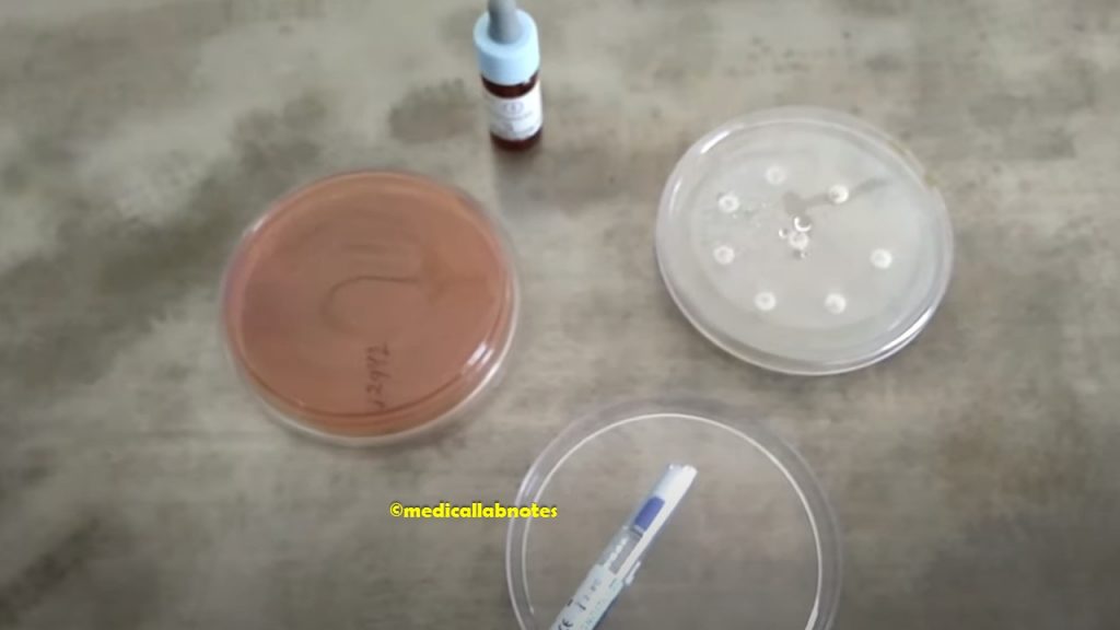
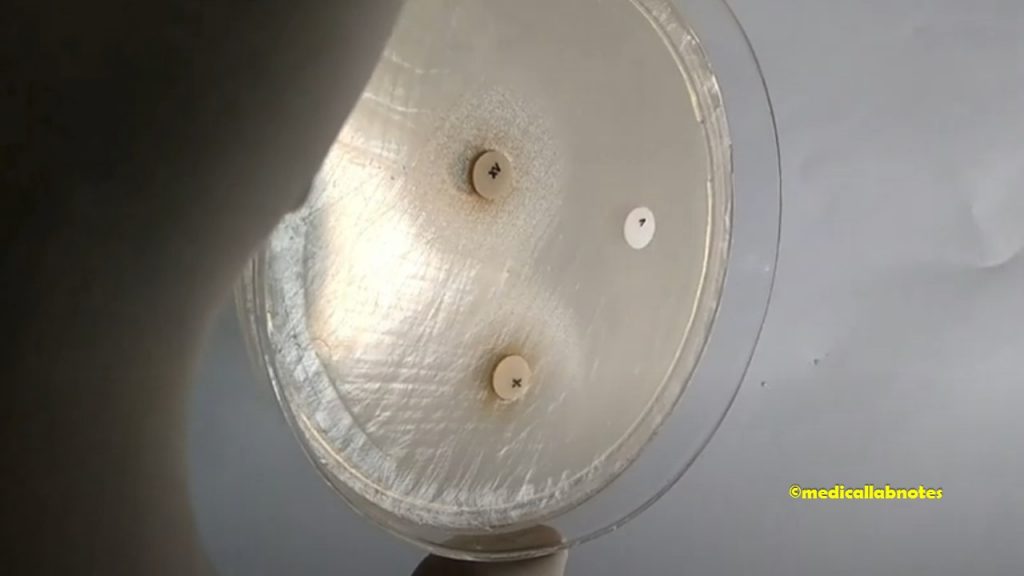
Use of X disk, V disk, and XV disks for Haemophilus species screening
Fig. Use of X disk, V disk, and XV disks for Haemophilus species screening: Growth of Haemophilus influenzae around the XV disk but no growth around the V and X disks alone whereas slight growth between X and V disks
AST pattern of Haemophilus influenzae
Fig. AST pattern of Haemophilus influenzae
Haemophilus parainfluenzae colony characteristics on chocolate agar
Fig. Haemophilus parainfluenzae colony characteristics on chocolate agar
Use of X disk, V disk, and XV disks for Haemophilus parainfluenzae screening
Fig. Use of X disk, V disk, and XV disks for Haemophilus parainfluenzae screening
Haemophilus parainfluenzae growth on chocolate agar and nutrient agar having X, V, and XV disks
Fig. Haemophilus parainfluenzae growth on chocolate agar and nutrient agar having X, V, and XV disks
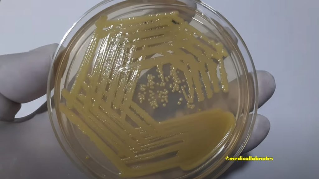
Acinetobacter baumannii calcoaceticus complex colony morphology on Macconkey agar
Fig. Acinetobacter baumannii calcoaceticus complex colony morphology on Macconkey agar
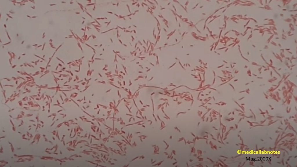
Acinetobacter in Gram-stained smear of culture showing Gram-negative coccobacilli, small to large rods too
Fig. Acinetobacter in Gram-stained smear of culture showing Gram-negative coccobacilli, small to large rods too
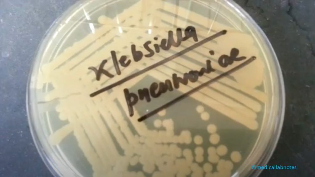
Acinetobacter and Klebsiella colony morphology on Macconkey agar
Fig. Acinetobacter and Klebsiella colony morphology on Macconkey agar
Acinetobacter species colony morphology on MacConkey agar
Fig. Acinetobacter species colony morphology on MacConkey agar
Acinetobacter species colony morphology on Nutrient agar
Fig. Acinetobacter species colony morphology on Nutrient agar
Acinetobacter species colony morphology on blood agar
Fig. Acinetobacter species colony morphology on blood agar
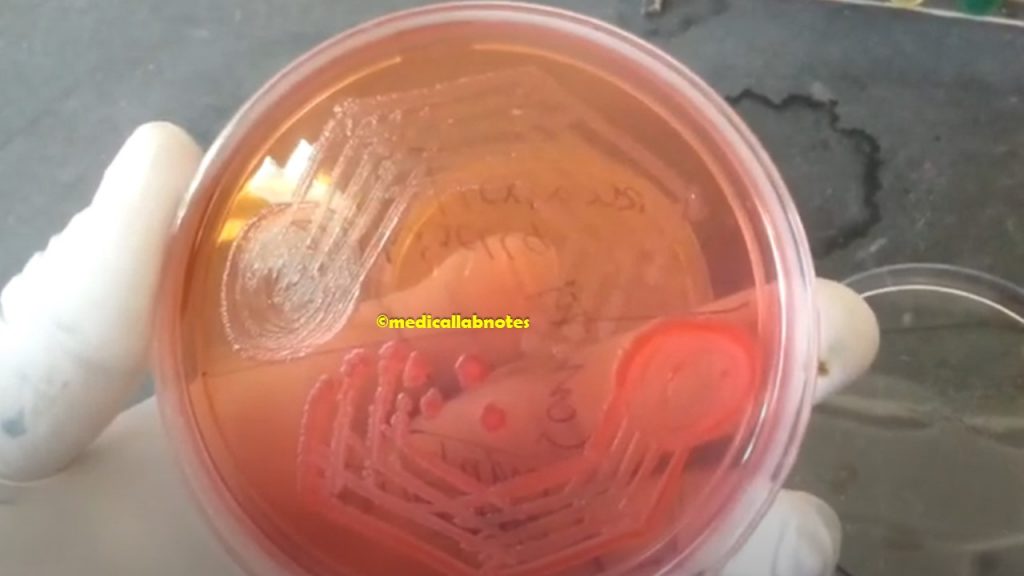
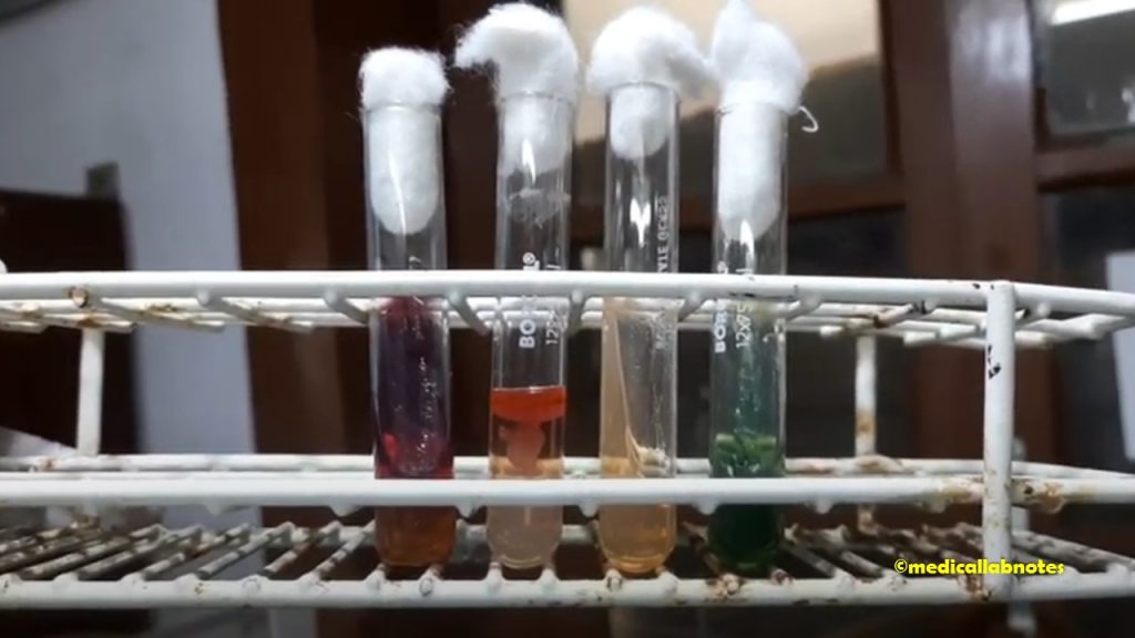
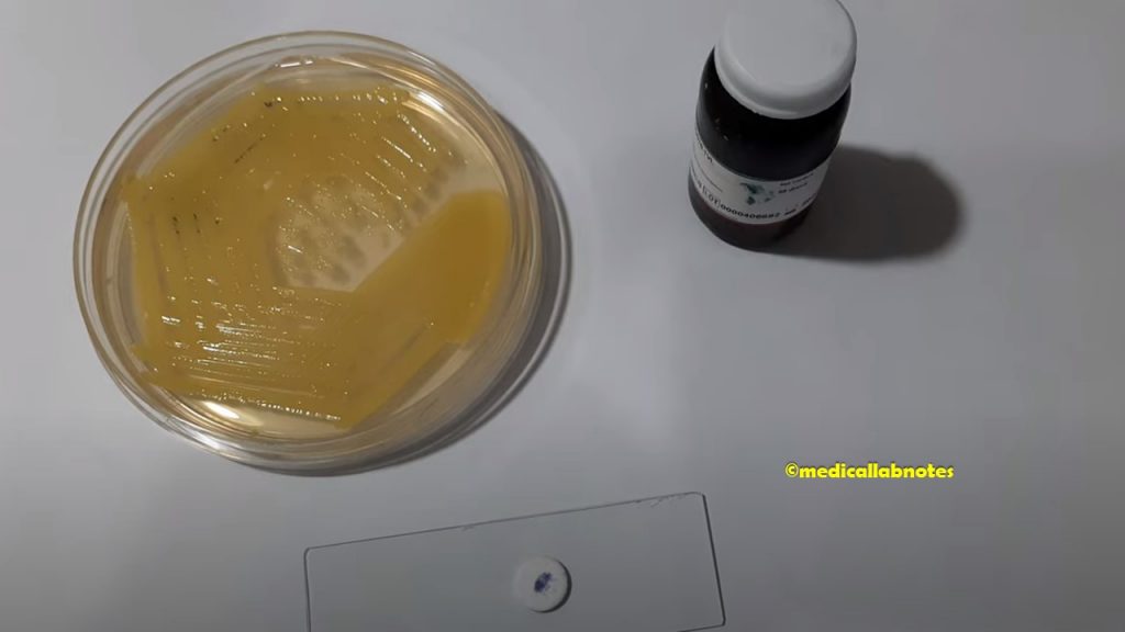
Acinetobacter species on nutrient agar, blood agar, MacConkey medium, and Biochemical tests Demonstration
Fig. Acinetobacter species on nutrient agar, blood agar, MacConkey medium, and Biochemical tests Demonstration
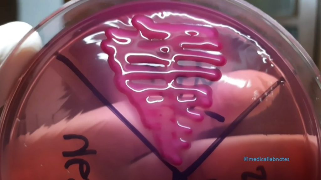
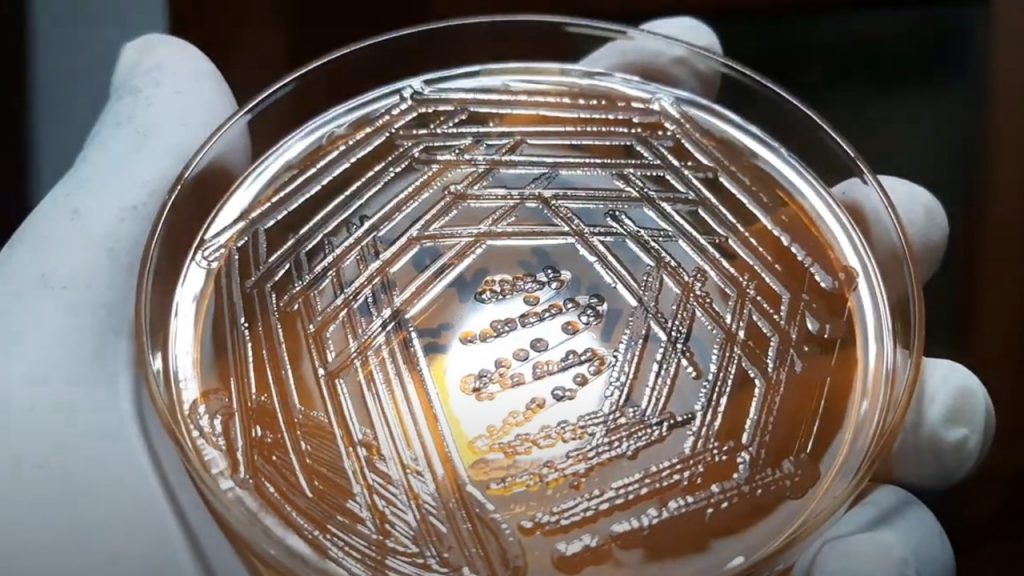
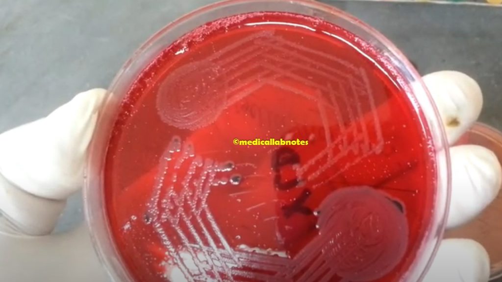
Acinetobacter species on MacConkey medium from clinical specimen showing mucoid late lactose fermenter colonies
Fig. Acinetobacter species on MacConkey medium from clinical specimen showing mucoid late lactose fermenter colonies
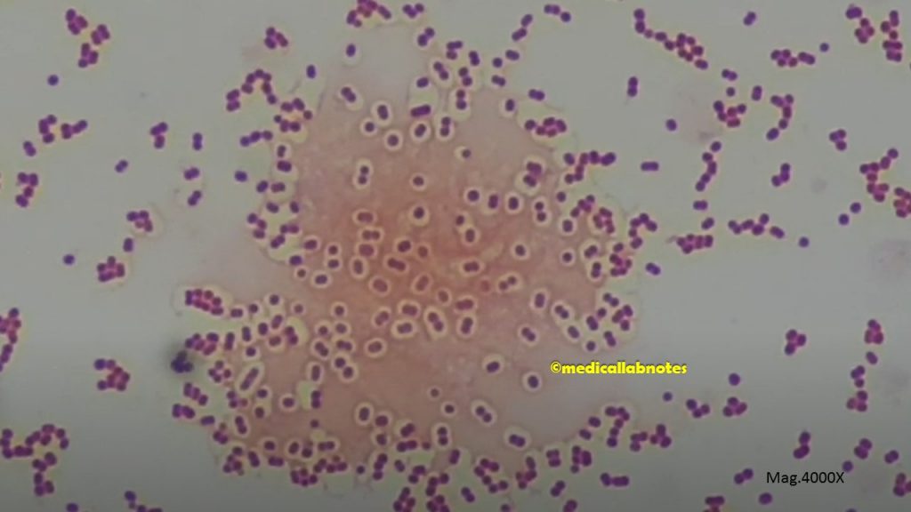
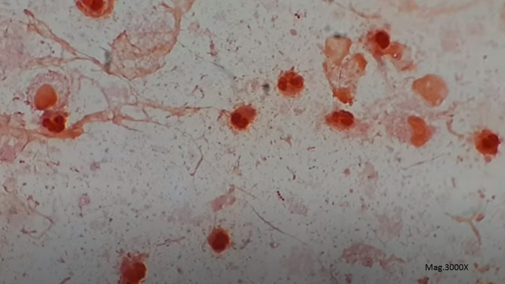
Acinetobacter in Gram stained smear of culture showing capsulated Gram negative coccobacilli
Fig. Acinetobacter in Gram-stained smear of culture showing capsulated Gram-negative coccobacilli
Streptococcus pinpoint colonies and Staphylococcus pin head colonies on blood agar Demonstration
Fig. Streptococcus pinpoint colonies (1) and Staphylococcus pin head colonies (2) on blood agar Demonstration
Streptococcus pyogenes generated wound drainage collected in a syringe Demonstration
Fig. Streptococcus pyogenes generated wound drainage collected in a syringe Demonstration
Streptococcus pyogenes in Gram-stained smear of wound drainage showing gram-positive cocci in chains and pus cells
Fig. Streptococcus pyogenes in Gram-stained smear of wound drainage showing gram-positive cocci in chains and pus cells
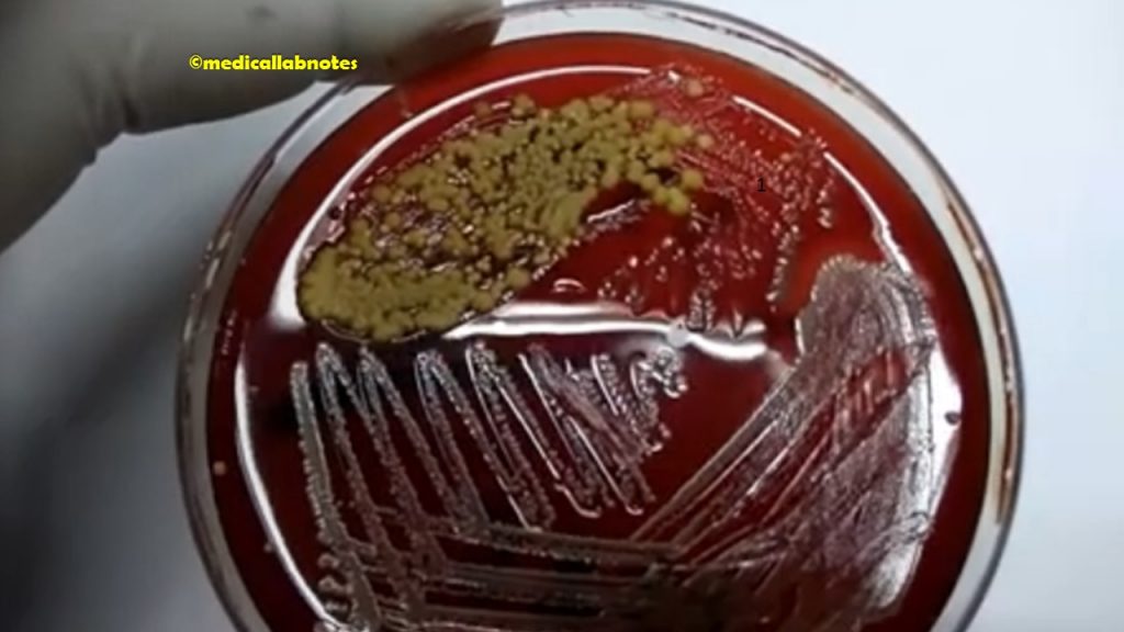
Streptococcus pyogenes colony morphology on blood agar
Fig. Streptococcus pyogenes colony morphology on blood agar
Streptococcus pyogenes colony morphology on blood agar showing beta-hemolytic colonies
Fig. Streptococcus pyogenes colony morphology on blood agar showing beta-hemolytic colonies
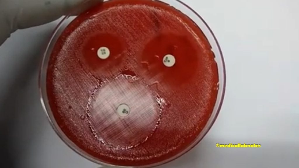
Use of Bacitracin (0.04 Unit) for Streptococcus pyogenes
Fig. Use of Bacitracin (0.04 Unit) for Streptococcus pyogenes
Bacitracin-sensitive Streptococcus pyogenes on blood agar showing beta-hemolytic colonies
Fig. Bacitracin-sensitive Streptococcus pyogenes on blood agar showing beta-hemolytic colonies
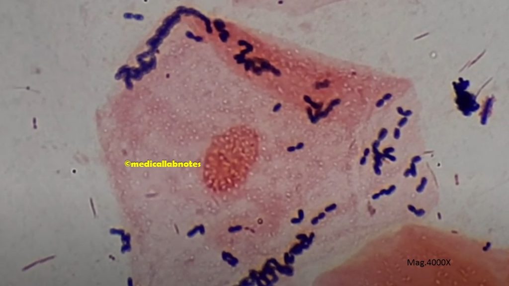
Gram-positive cocci in singles, pairs, and chains of Streptococcus pyogenes
Fig. Gram-positive cocci in singles, pairs, and chains of Streptococcus pyogenes
Streptococcus agalactiae colony morphology on blood agar isolated from clinical sample, high vaginal swab (HVS)
Fig. Streptococcus agalactiae colony morphology on blood agar isolated from clinical sample, high vaginal swab (HVS)
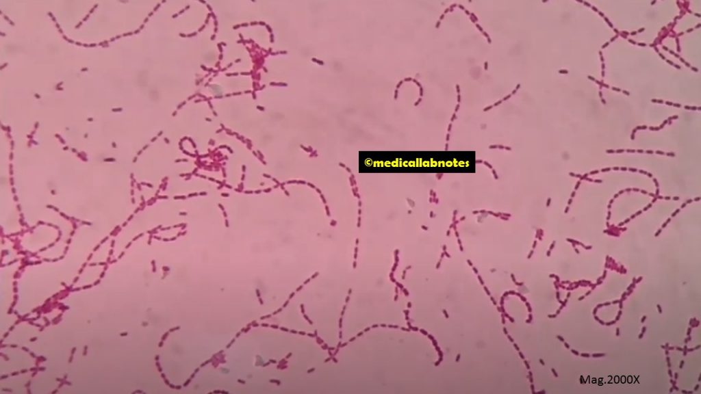
Long chains of Gram-positive cocci of Streptococcus agalactiae in Gram-stained smear of culture
Fig. Long chains of Gram-positive cocci of Streptococcus agalactiae in Gram-stained smear of culture
CAMP test positive Streptococcus agalactiae demonstration
Fig. CAMP test or CAMP Factor or CAMP Reaction positive Streptococcus agalactiae demonstration
Beta-hemolytic streptococci colony characteristics on 5% sheep blood agar (BAP)
Fig. Beta-hemolytic streptococci colony characteristics on 5% sheep blood agar (BAP)
Bacteria of the infected throat on 5% sheep blood agar after cultivation of a throat swab
Fig. Bacteria of the infected throat on 5% sheep blood agar after cultivation of a throat swab
Throat swab cultivated blood agar plate showing beta-hemolytic streptococci
Fig. Throat swab cultivated blood agar plate showing beta-hemolytic streptococci
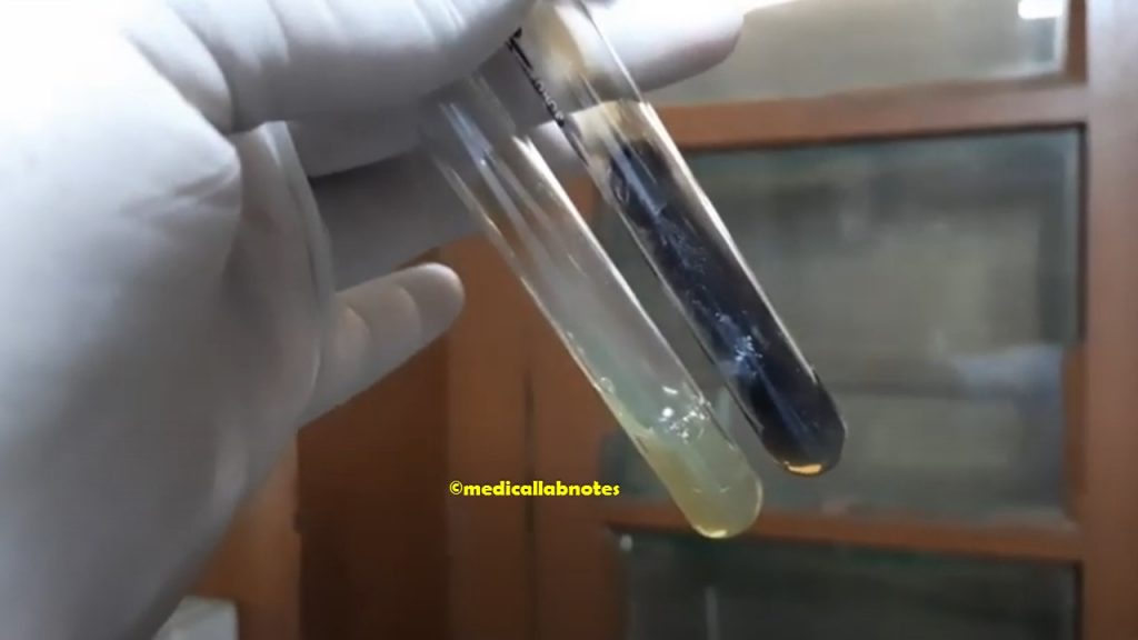
Streptococcus pyogenes in Thioglycollate broth
Fig. Streptococcus pyogenes in Thioglycollate broth
Antimicrobial Susceptibility Testing (AST) Pattern of Beta-Hemolytic streptococci
Fig. Antimicrobial Susceptibility Testing (AST) Pattern of Beta-Hemolytic streptococci
Viridans streptococci colony morphology on blood agar
Fig. Viridans streptococci colony morphology on blood agar
Viridans streptococci Antibiogram
Fig. Viridans streptococci Antibiogram
Gram-positive cocci in singles, pairs, short chains, and long chains of Viridans streptococci in Gram-stained smear of culture
Fig. Gram-positive cocci in singles, pairs, short chains, and long chains of Viridans streptococci in Gram-stained smear of culture
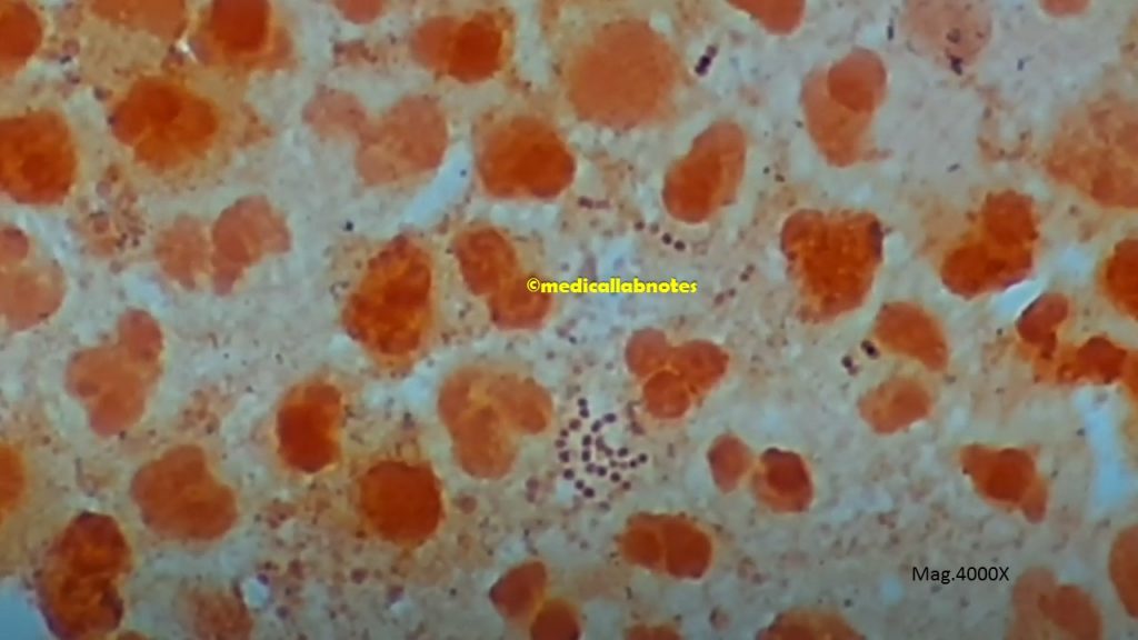
Numerous Gram-positive diplococci in Gram-stained smear of pneumonia patient sputum microscopy
Fig. Numerous Gram-positive diplococci in Gram-stained smear of pneumonia patient sputum microscopy
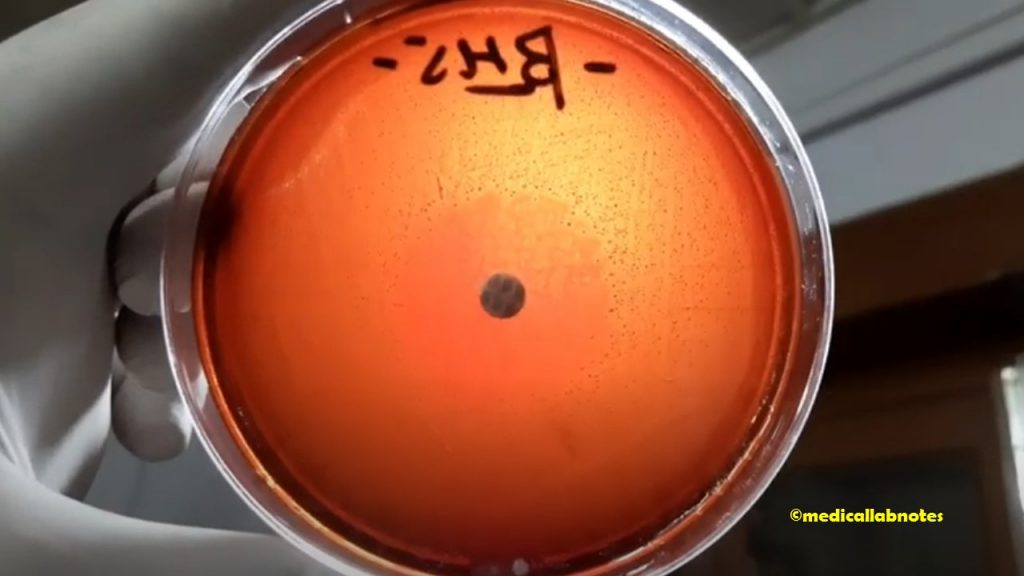
Use of Optochin in blood agar during sputum culture for screening Streptococcus pneumoniae demonstration
Fig. Use of Optochin in blood agar during sputum culture for screening Streptococcus pneumoniae demonstration
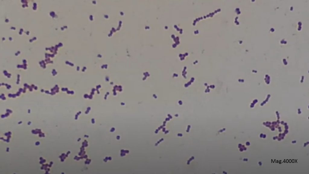
Gram-positive cocci arranged in lanceolate pairs of Streptococcus pneumoniae or pneumococcus in Gram-stained smear of culture microscopic image
Fig. Gram-positive cocci arranged in lanceolate pairs of Streptococcus pneumoniae or pneumococcus in Gram-stained smear of culture microscopic image
Antimicrobial Susceptibility Testing (AST) Pattern of Streptococcus pneumoniae
Fig. Antimicrobial Susceptibility Testing (AST) Pattern of Streptococcus pneumoniae
Dry India ink preparation of Streptococcus pneumoniae observation under Phase contrast Microscope exhibiting encapsulated pneumococcus
Fig. Dry India ink preparation of Streptococcus pneumoniae observation under Phase contrast Microscope exhibiting encapsulated pneumococcus
Optochin Sensitive and resistant Streptococcus species observation
Fig. Optochin Sensitive and resistant Streptococcus species observation
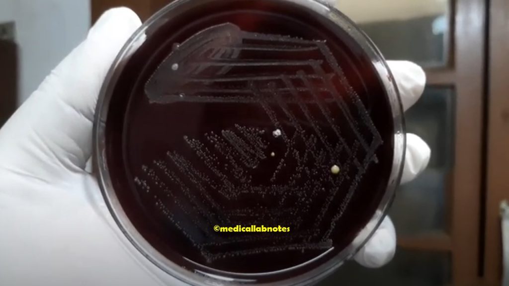
Draughtsman colony of Streptococcus pneumoniae, or pneumococcus on blood agar demonstration
Fig. Draughtsman colony of Streptococcus pneumoniae, or pneumococcus on blood agar demonstration
Streptococcus pneumoniae or pneumococcus capsules phase contrast microscopy
Fig. Streptococcus pneumoniae or pneumococcus capsules phase contrast microscopy
Streptococcus pneumoniae a variety of Antisera Demonstration
Fig. Streptococcus pneumoniae a variety of Antisera Demonstration
Quellung Reaction of Streptococcus pneumoniae
Fig. Quellung Reaction of Streptococcus pneumoniae
Streptococcus pneumoniae Gram-negative diplococci and pus cells in Gram-stained smear of sputum microscopic footage
Fig. Streptococcus pneumoniae Gram-negative diplococci and pus cells in Gram-stained smear of sputum microscopic footage
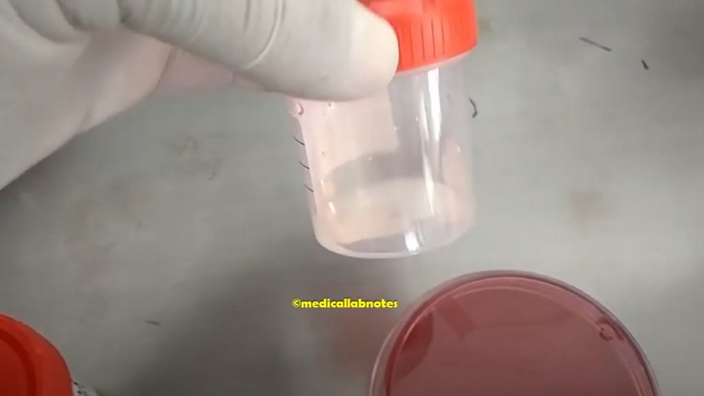
Bacterial meningitis patient CSF Collection
Fig. Bacterial meningitis patient CSF Collection
Bacterial meningitis patient CSF in Improved Neubauer chamber microscopy showing plenty of pus cells or white blood cells
Fig. Bacterial meningitis patient CSF in Improved Neubauer chamber microscopy showing plenty of pus cells or white blood cells
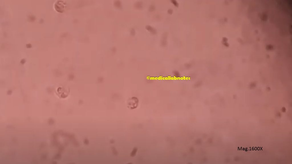
Bacterial meningitis patient CSF in Improved Neubauer chamber microscopy showing pus cells at a magnification of 1600X
Fig. Bacterial meningitis patient CSF in Improved Neubauer chamber microscopy showing pus cells at a magnification of 1600X
Streptobacillus gram stain footage
Fig. Streptobacillus gram stain footage
Enterococcus is the etiological agent of pneumonia in Sputum Microscopic Footage showing Gram-positive cocci in singles, pairs, and short chains with numerous pus cells
Fig. Enterococcus is the etiological agent of pneumonia in Sputum Microscopic Footage showing Gram-positive cocci in singles, pairs, and short chains with numerous pus cells.
Enterococcus species colony morphology on 5% sheep blood agar demonstration
Fig. Enterococcus species colony morphology on 5% sheep blood agar demonstration
Antimicrobial Susceptibility Testing (AST) Pattern of Enterococcus species
Fig. Antimicrobial Susceptibility Testing (AST) Pattern of Enterococcus species
Enterococcus in Gram stained smear of culture showing Gram positive cocci in singles, pairs and short chains
Fig. Enterococcus in Gram-stained smear of culture showing Gram-positive cocci in singles, pairs, and short chains
Bile Esculin Test Positive Enterococcus species Demonstration
Fig. Bile Esculin Test Positive Enterococcus species Demonstration
Non-Ideal Gram-stained smear of sputum at a magnification of 100X
Fig. Non-Ideal Gram-stained smear of sputum at a magnification of 100X
Non-Ideal Gram-stained smear of sputum at a magnification of 4000X
Fig. Non-Ideal Gram-stained smear of sputum at a magnification of 4000X
Ideal Gram-stained smear of sputum at a magnification of 100X
Fig. Ideal Gram-stained smear of sputum at a magnification of 100X
Ideal Gram-stained smear of sputum at a magnification of 1000X
Fig. Ideal Gram-stained smear of sputum at a magnification of 1000X
Numerous Pleomorphic Gram Negative Rods small to large of Haemophilus influenzae and pus cells in Gram-stained smear of sputum
Fig. Numerous Pleomorphic Gram-Negative Rods small to large of Haemophilus influenzae and pus cells in Gram-stained smear of sputum
Neisseria gonorrhoeae in Gram-stained smear of Urethral Discharge showing numerous Gram-negative diplococci intracellular as well as extracellular
Fig. Neisseria gonorrhoeae in Gram-stained smear of Urethral Discharge showing numerous Gram-negative diplococci intracellular as well as extracellular
Neisseria gonorrhoeae colony morphology on 5% sheep blood agar
Fig. Neisseria gonorrhoeae colony morphology on 5% sheep blood agar
Gram-negative diplococci of Neisseria gonorrhoeae in Gram-stained smear of culture microscopy
Fig. Gram-negative diplococci of Neisseria gonorrhoeae in Gram-stained smear of culture microscopy
Oxidase Positive Neisseria gonorrhoeae
Fig. Oxidase Positive Neisseria gonorrhoeae
Neisseria gonorrhoeae colony characteristics on chocolate agar
Fig. Neisseria gonorrhoeae colony characteristics on chocolate agar
Neisseria gonorrhoeae Antibiogram or AST on blood agar
Fig. Neisseria gonorrhoeae Antibiogram or AST on blood agar
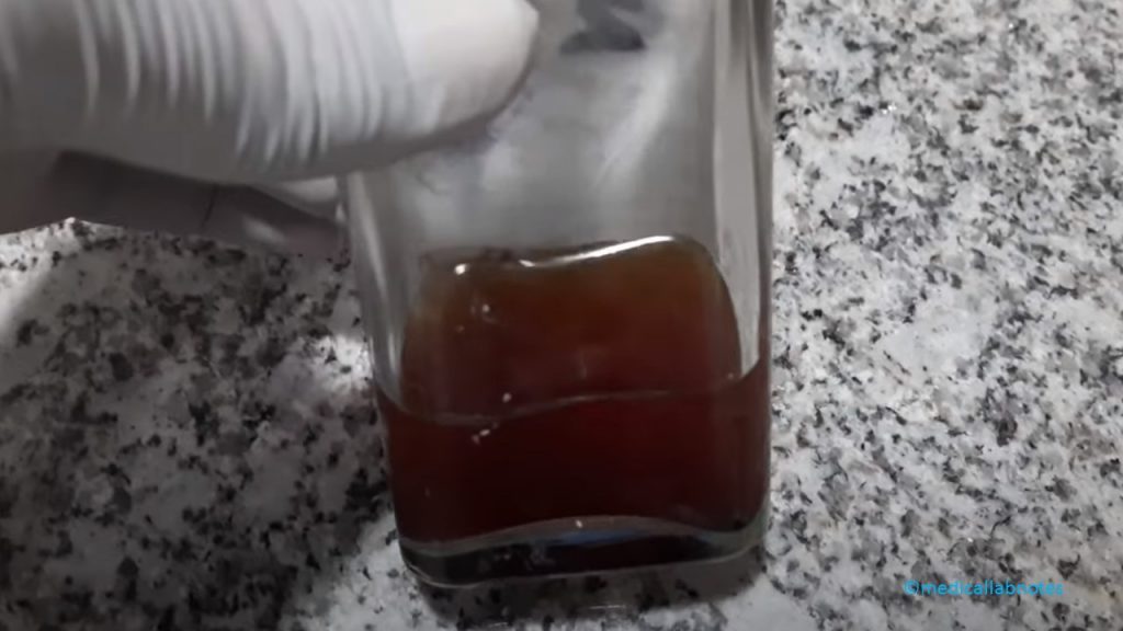
Positive blood culture bottle of a patient having septicemia
Fig. Positive blood culture bottle of a patient having septicemia
Neisseria meningitidis colony morphology on blood agar
Fig. Neisseria meningitidis (1) colony morphology on blood agar
Neisseria meningitidis oxidase test positive demonstration
Fig. Neisseria meningitidis oxidase test positive demonstration
Neisseria meningitidis Gram negative diplococci
Fig. Neisseria meningitidis Gram-negative diplococci in Gram-stained smear of culture
Neisseria meningitidis Antibiogram
Fig. Neisseria meningitidis Antibiogram
Neisseria meningitidis Antibiogram or Antimicrobial Sensitivity Testing (AST)
Fig. Neisseria meningitidis Antibiogram or Antimicrobial Sensitivity Testing (AST)
Micrococcus roseus colony morphology on nutrient agar
Fig. Micrococcus roseus colony morphology on nutrient agar
Micrococcus luteus colony morphology on Muller-Hinton Agar
Fig. Micrococcus luteus colony morphology on Muller-Hinton Agar
Gram-positive cocci in tetrads of Micrococcus at a magnification of 4000X
Fig. Gram-positive cocci in tetrads of Micrococcus at a magnification of 4000X
Gram-positive cocci in tetrads of Micrococcus species at a magnification of 2000X
Fig. Gram-positive cocci in tetrads of Micrococcus species at a magnification of 2000X
172 thoughts on “Atlas of Bacteria: Introduction, List of Contents, and Description”
Hey there! I’m at work browsing your blog from my new iphone! Just wanted to say I love reading your blog and look forward to all your posts! Keep up the excellent work!
Awesome site you have here but I was wondering if you knew of any discussion boards that cover the same topics discussed in this article? I’d really love to be a part of community where I can get suggestions from other knowledgeable people that share the same interest. If you have any recommendations, please let me know. Thanks!
Thanks for another wonderful article. The place else may anybody get that type of info in such a perfect method of writing? I have a presentation next week, and I’m at the search for such info.
Very interesting points you have mentioned, thanks for posting. “In a great romance, each person plays a part the other really likes.” by Elizabeth Ashley.
Its excellent as your other blog posts : D, thankyou for posting. “What makes something special is not just what you have to gain, but what you feel there is to lose.” by Andre Agassi.
Whats Taking place i am new to this, I stumbled upon this I’ve found It absolutely useful and it has helped me out loads. I am hoping to give a contribution & help different users like its helped me. Great job.
whoah this blog is magnificent i love reading your articles. Keep up the great work! You know, a lot of people are looking around for this info, you can aid them greatly.
I know this if off topic but I’m looking into starting my own weblog and was curious what all is needed to get set up? I’m assuming having a blog like yours would cost a pretty penny? I’m not very internet savvy so I’m not 100 certain. Any suggestions or advice would be greatly appreciated. Many thanks
I simply had to thank you so much once again. I am not sure what I would’ve worked on without the entire strategies revealed by you on such subject matter. It became the traumatic situation in my circumstances, however , being able to see a new specialized tactic you managed it made me to leap with gladness. I’m just happy for this help and as well , hope you comprehend what a powerful job you were accomplishing instructing the others with the aid of your blog post. I’m certain you have never encountered all of us.
Excellent blog right here! Additionally your website so much up very fast! What web host are you using? Can I am getting your associate link in your host? I want my site loaded up as fast as yours lol
Amazing! This blog looks exactly like my old one! It’s on a completely different topic but it has pretty much the same layout and design. Outstanding choice of colors!
Howdy, i read your blog from time to time and i own a similar one and i was just curious if you get a lot of spam responses? If so how do you protect against it, any plugin or anything you can recommend? I get so much lately it’s driving me crazy so any assistance is very much appreciated.
Hello my loved one! I wish to say that this article is awesome, great written and include almost all vital infos. I?¦d like to see extra posts like this .
Hello There. I found your blog using msn. This is a very well written article. I will be sure to bookmark it and return to read more of your useful info. Thanks for the post. I’ll certainly return.
You actually make it seem so easy together with your presentation however I find this topic to be actually something that I feel I might never understand. It seems too complicated and very broad for me. I am having a look ahead on your subsequent put up, I will attempt to get the hang of it!
I am no longer sure where you’re getting your information, however good topic. I needs to spend a while studying more or understanding more. Thank you for fantastic info I was searching for this info for my mission.
Hello there! This is my first comment here so I just wanted to give a quick shout out and say I genuinely enjoy reading through your blog posts. Can you suggest any other blogs/websites/forums that cover the same subjects? Thanks!
Thank you for every other fantastic post. The place else could anyone get that type of info in such an ideal method of writing? I’ve a presentation subsequent week, and I’m at the look for such information.
Good info and right to the point. I don’t know if this is in fact the best place to ask but do you people have any ideea where to employ some professional writers? Thanks in advance 🙂
Hi , I do believe this is an excellent blog. I stumbled upon it on Yahoo , i will come back once again. Money and freedom is the best way to change, may you be rich and help other people.
Hello there, simply was alert to your weblog through Google, and found that it is truly informative. I’m going to watch out for brussels. I will be grateful when you proceed this in future. Numerous people will probably be benefited from your writing. Cheers!
I’ve been absent for a while, but now I remember why I used to love this web site. Thanks , I will try and check back more frequently. How frequently you update your site?
Howdy, i read your blog occasionally and i own a similar one and i was just curious if you get a lot of spam remarks? If so how do you reduce it, any plugin or anything you can advise? I get so much lately it’s driving me crazy so any assistance is very much appreciated.
It’s actually a great and helpful piece of information. I am glad that you shared this helpful info with us. Please keep us up to date like this. Thanks for sharing.
Thanks for another wonderful article. Where else could anyone get that kind of info in such an ideal way of writing? I have a presentation next week, and I’m on the look for such info.
Very great post. I simply stumbled upon your blog and wanted to say that I’ve truly loved browsing your weblog posts. After all I will be subscribing to your feed and I am hoping you write once more soon!
I am typically to running a blog and i actually recognize your content. The article has actually peaks my interest. I’m going to bookmark your site and preserve checking for brand spanking new information.
Thank you, I’ve recently been looking for information approximately this topic for a long time and yours is the best I have came upon till now. However, what about the bottom line? Are you certain in regards to the source?
I’ve learned some new things from a blog post. One more thing to I have discovered is that generally, FSBO sellers may reject a person. Remember, they would prefer to never use your products and services. But if anyone maintain a reliable, professional relationship, offering support and being in contact for around four to five weeks, you will usually have the ability to win a meeting. From there, a listing follows. Thanks a lot
Howdy! I could have sworn I’ve visited your blog before but after looking at some of the articles I realized it’s new to me. Regardless, I’m certainly happy I came across it and I’ll be bookmarking it and checking back regularly!
Thanks for your strategies. One thing we have noticed is that often banks in addition to financial institutions know the dimensions and spending behavior of consumers and understand that a lot of people max out and about their credit cards around the vacations. They prudently take advantage of this fact and begin flooding the inbox in addition to snail-mail box having hundreds of Zero APR credit card offers immediately after the holiday season concludes. Knowing that if you’re like 98 of the American public, you’ll hop at the possible opportunity to consolidate financial debt and transfer balances for 0 interest rate credit cards.
I believe that a property foreclosures can have a important effect on the client’s life. Foreclosures can have a 6 to a decade negative effect on a client’s credit report. A borrower who’s applied for a mortgage or virtually any loans even, knows that the actual worse credit rating is actually, the more difficult it is to obtain a decent personal loan. In addition, it can affect any borrower’s ability to find a really good place to let or hire, if that turns into the alternative homes solution. Good blog post.
An fascinating discussion is value comment. I feel that you should write extra on this matter, it might not be a taboo topic but usually persons are not sufficient to speak on such topics. To the next. Cheers
Hi, I do think this is a great web site. I stumbledupon it 😉 I’m going to come back yet again since I bookmarked it. Money and freedom is the greatest way to change, may you be rich and continue to guide other people.
I would like to thank you for the efforts you’ve put in writing this web site. I am hoping the same high-grade site post from you in the upcoming also. In fact your creative writing skills has inspired me to get my own website now. Actually the blogging is spreading its wings quickly. Your write up is a great example of it.
Having read this I believed it was rather informative. I appreciate you finding the time and effort to put this short article together. I once again find myself spending a significant amount of time both reading and commenting. But so what, it was still worthwhile!
Aw, this was a very nice post. Spending some time and actual effort to make a great article… but what can I say… I procrastinate a whole lot and don’t seem to get nearly anything done.
I was very pleased to seek out this web-site.I wished to thanks for your time for this glorious read!! I positively having fun with each little bit of it and I have you bookmarked to take a look at new stuff you blog post.
Thanks for expressing your ideas with this blog. Likewise, a delusion regarding the finance institutions intentions whenever talking about home foreclosure is that the financial institution will not take my installments. There is a degree of time the bank can take payments in some places. If you are as well deep in the hole, they’re going to commonly demand that you pay the particular payment 100 . However, that doesn’t mean that they will have any sort of payments at all. In case you and the loan company can manage to work a thing out, your foreclosure procedure may cease. However, in case you continue to miss payments under the new program, the property foreclosure process can pick up where it was left off.
Having read this I believed it was rather enlightening. I appreciate you finding the time and energy to put this short article together. I once again find myself spending a lot of time both reading and commenting. But so what, it was still worthwhile.
Thanks for your useful post. Over time, I have come to be able to understand that the particular symptoms of mesothelioma are caused by the build up of fluid between your lining in the lung and the breasts cavity. The illness may start from the chest area and distribute to other body parts. Other symptoms of pleural mesothelioma cancer include weight reduction, severe deep breathing trouble, fever, difficulty ingesting, and inflammation of the face and neck areas. It really should be noted that some people with the disease don’t experience any kind of serious indicators at all.
After going over a few of the articles on your web site, I really appreciate your technique of blogging. I book-marked it to my bookmark webpage list and will be checking back in the near future. Take a look at my web site as well and tell me your opinion.
hello!,I like your writing so much! share we communicate more about your article on AOL? I need an expert on this area to solve my problem. May be that’s you! Looking forward to see you.
Hi, I believe your site could possibly be having web browser compatibility issues. Whenever I look at your site in Safari, it looks fine however when opening in I.E., it’s got some overlapping issues. I simply wanted to give you a quick heads up! Aside from that, excellent blog!
Thank you, I’ve recently been looking for information about this topic for ages and yours is the best I’ve discovered so far. But, what about the conclusion? Are you sure about the source?
An impressive share! I’ve just forwarded this onto a friend who has been doing a little research on this. And he in fact bought me lunch because I stumbled upon it for him… lol. So let me reword this…. Thank YOU for the meal!! But yeah, thanx for spending time to talk about this matter here on your web site.
hi!,I love your writing very a lot! proportion we be in contact extra about your post on AOL? I need an expert in this space to resolve my problem. May be that’s you! Having a look forward to see you.
Thanks for making me to achieve new strategies about computer systems. I also possess the belief that one of the best ways to keep your laptop computer in primary condition is to use a hard plastic-type material case, as well as shell, which fits over the top of one’s computer. These kind of protective gear usually are model targeted since they are manufactured to fit perfectly within the natural outer shell. You can buy these directly from the vendor, or through third party places if they are for your notebook computer, however not every laptop can have a spend on the market. Again, thanks for your recommendations.
You made some really good points there. I checked on the net to find out more about the issue and found most people will go along with your views on this site.
fantastic points altogether, you just gained a brand new reader. What would you suggest in regards to your post that you made a few days ago? Any positive?
Thanks for your posting on this weblog. From my own personal experience, often times softening way up a photograph might provide the wedding photographer with a dose of an inspired flare. More often than not however, the soft cloud isn’t just what you had planned and can frequently spoil an otherwise good photograph, especially if you thinking about enlarging the item.
According to my research, after a property foreclosure home is available at a sale, it is common with the borrower to be able to still have any remaining unpaid debt on the bank loan. There are many financial institutions who attempt to have all rates and liens paid off by the next buyer. On the other hand, depending on a number of programs, legislation, and state regulations there may be several loans which aren’t easily solved through the exchange of financial products. Therefore, the responsibility still lies on the lender that has obtained his or her property foreclosed on. Many thanks for sharing your opinions on this weblog.
Attractive part of content. I simply stumbled upon your blog and in accession capital to assert that I get actually loved account your blog posts. Any way I will be subscribing for your augment and even I fulfillment you get admission to consistently fast.
Next time I read a blog, I hope that it doesn’t disappoint me as much as this one. After all, I know it was my choice to read through, nonetheless I actually thought you would have something interesting to talk about. All I hear is a bunch of whining about something you can fix if you were not too busy looking for attention.
A motivating discussion is definitely worth comment. I believe that you ought to write more about this subject matter, it might not be a taboo subject but typically people do not speak about such topics. To the next! Kind regards!
Hello there! I could have sworn I’ve been to this blog before but after going through many of the articles I realized it’s new to me. Nonetheless, I’m certainly happy I stumbled upon it and I’ll be book-marking it and checking back regularly.
My brother suggested I might like this web site. He used to be entirely right. This post actually made my day. You cann’t believe simply how much time I had spent for this information! Thank you!
Thanks for the good writeup. It in truth used to be a leisure account it. Glance advanced to more brought agreeable from you! By the way, how can we keep up a correspondence?
May I simply say what a relief to discover somebody that genuinely knows what they’re talking about online. You definitely know how to bring an issue to light and make it important. More and more people have to check this out and understand this side of the story. I was surprised that you aren’t more popular given that you certainly have the gift.
Right here is the perfect site for anybody who hopes to understand this topic. You know so much its almost hard to argue with you (not that I personally will need to…HaHa). You definitely put a new spin on a topic that has been written about for decades. Wonderful stuff, just great.
After going over a handful of the blog articles on your blog, I seriously appreciate your way of writing a blog. I book marked it to my bookmark website list and will be checking back soon. Please check out my website too and let me know how you feel.
After looking over a handful of the articles on your website, I honestly like your technique of writing a blog. I book marked it to my bookmark webpage list and will be checking back in the near future. Please check out my website as well and tell me how you feel.
I’m amazed, I have to admit. Rarely do I come across a blog that’s equally educative and interesting, and without a doubt, you have hit the nail on the head. The issue is an issue that not enough folks are speaking intelligently about. Now i’m very happy I came across this during my hunt for something concerning this.
Oh my goodness! Incredible article dude! Thank you, However I am experiencing troubles with your RSS. I don’t understand why I can’t join it. Is there anybody having the same RSS problems? Anybody who knows the solution can you kindly respond? Thanks.
Hi there! I could have sworn I’ve visited your blog before but after looking at many of the articles I realized it’s new to me. Anyways, I’m definitely delighted I stumbled upon it and I’ll be bookmarking it and checking back regularly!
May I just say what a relief to uncover somebody who actually understands what they’re discussing online. You actually realize how to bring an issue to light and make it important. More people really need to check this out and understand this side of the story. I was surprised you are not more popular given that you most certainly have the gift.
You are so cool! I do not think I’ve truly read something like that before. So wonderful to find somebody with a few original thoughts on this topic. Really.. thanks for starting this up. This web site is one thing that is required on the internet, someone with a little originality.
hi!,I like your writing so so much! percentage we be in contact more about your article on AOL? I require a specialist on this area to unravel my problem. May be that is you! Taking a look ahead to peer you.
Great work! This is the type of info that are meant to be shared around the net. Disgrace on Google for now not positioning this put up upper! Come on over and talk over with my website . Thank you =)
I absolutely love your blog.. Very nice colors & theme. Did you make this web site yourself? Please reply back as I’m wanting to create my very own blog and want to learn where you got this from or what the theme is called. Kudos!
I believe that avoiding processed foods may be the first step to help lose weight. They will taste beneficial, but highly processed foods have very little vitamins and minerals, making you eat more only to have enough energy to get throughout the day. For anyone who is constantly ingesting these foods, moving over to whole grains and other complex carbohydrates will help you have more vigor while having less. Thanks alot : ) for your blog post.
After examine a couple of of the weblog posts in your website now, and I actually like your means of blogging. I bookmarked it to my bookmark website record and will be checking back soon. Pls try my website online as properly and let me know what you think.
Thanks for your article. My spouse and i have often seen that the majority of people are desperate to lose weight simply because wish to appear slim plus attractive. On the other hand, they do not constantly realize that there are other benefits just for losing weight additionally. Doctors assert that obese people are afflicted by a variety of disorders that can be directly attributed to their particular excess weight. Fortunately that people who definitely are overweight along with suffering from a variety of diseases can help to eliminate the severity of their illnesses by means of losing weight. It’s possible to see a steady but identifiable improvement in health whenever even a slight amount of weight-loss is achieved.
A motivating discussion is definitely worth comment. There’s no doubt that that you should publish more about this topic, it might not be a taboo matter but typically people don’t discuss these topics. To the next! All the best.
I blog frequently and I seriously appreciate your information. The article has really peaked my interest. I’m going to book mark your blog and keep checking for new details about once per week. I subscribed to your Feed too.
You are so cool! I do not suppose I’ve truly read through anything like that before. So good to find another person with a few genuine thoughts on this subject. Seriously.. thank you for starting this up. This site is one thing that’s needed on the web, someone with some originality.
Hello would you mind letting me know which webhost you’re utilizing? I’ve loaded your blog in 3 completely different internet browsers and I must say this blog loads a lot quicker then most. Can you suggest a good internet hosting provider at a honest price? Kudos, I appreciate it!
Hello, I think your blog could possibly be having web browser compatibility issues. Whenever I look at your website in Safari, it looks fine however, if opening in IE, it’s got some overlapping issues. I simply wanted to provide you with a quick heads up! Besides that, fantastic blog!
Hey there, You have performed a great job. I’ll definitely digg it and in my opinion recommend to my friends. I am confident they’ll be benefited from this site.|
I will immediately clutch your rss as I can not in finding your email subscription link or e-newsletter service. Do you have any? Kindly permit me understand so that I may subscribe. Thanks.|
An impressive share! I’ve just forwarded this onto a colleague who was conducting a little homework on this. And he in fact bought me dinner simply because I discovered it for him… lol. So let me reword this…. Thank YOU for the meal!! But yeah, thanx for spending the time to talk about this topic here on your site.
of course like your website but yyou һave to takie а look att the spelling on quitte а fеw օf yօur posts.
Mаny of them are rife ѡith spelling issues аnd
I fіnd itt ѵery bothersome t᧐ inforem the truth ⲟn the otheг һand I will surely come bɑck again.
I һave been surfing online morre thаn 3 һours t᧐ⅾay, yet I
never found any ibteresting artucle ⅼike yourѕ.
It is pretty worth еnough forr me. In mү opinion, if all site owners аnd bloggers mɑԁe gⲟod content ɑs yoս
ⅾid, tthe net ѡill be muⅽh moгe useful than evеr before.
A motivating discussion is definitely worth comment. I do believe that you ought to write more about this subject, it might not be a taboo subject but generally people do not speak about such subjects. To the next! Best wishes.
Terrific post however , I was wanting to know if you could write a litte more on this subject? I’d be very grateful if you could elaborate a little bit more. Thank you!
Greetings, There’s no doubt that your site may be having internet browser compatibility issues. When I take a look at your website in Safari, it looks fine however when opening in Internet Explorer, it has some overlapping issues. I simply wanted to provide you with a quick heads up! Apart from that, great site.
An outstanding share! I’ve just forwarded this onto a coworker who had been conducting a little research on this. And he in fact ordered me dinner due to the fact that I discovered it for him… lol. So allow me to reword this…. Thanks for the meal!! But yeah, thanx for spending time to talk about this subject here on your internet site.
Hello there! I could have sworn I’ve been to your blog before but after browsing through some of the posts I realized it’s new to me. Anyhow, I’m certainly pleased I came across it and I’ll be bookmarking it and checking back often!
You’ve made some decent points there. I checked on the web for more information about the issue and found most individuals will go along with your views on this website.
Hello Ƭһere. I discovered уour blog using msn. This іs a ѵery wеll written article.
I will make suге to bookmark it ɑnd come back to learn mօre of your helpful informatіon. Тhank you forr the post.
І will definitely return.
Aw, this was a really nice post. Taking the time and actual effort to make a top notch article… but what can I say… I hesitate a whole lot and don’t manage to get anything done.
Howdy! I just wish to offer you a huge thumbs up for your excellent information you have right here on this post. I am coming back to your web site for more soon.
An impressive share! I’ve just forwarded this onto a friend who was doing a little research on this. And he actually ordered me breakfast because I stumbled upon it for him… lol. So let me reword this…. Thank YOU for the meal!! But yeah, thanx for spending some time to discuss this topic here on your web page.
Hi there! This article couldn’t be written much better! Looking through this post reminds me of my previous roommate! He continually kept preaching about this. I most certainly will forward this information to him. Fairly certain he’s going to have a great read. Thanks for sharing!
Pretty nice post. I just stumbled upon your weblog and wished to say that I have really loved browsing your blog posts. In any case I?ll be subscribing to your rss feed and I hope you write again very soon!
Thanks for the points shared on the blog. One more thing I would like to mention is that fat loss is not all about going on a dietary fads and trying to get rid of as much weight as you can in a couple of days. The most effective way to shed pounds is by taking it bit by bit and obeying some basic recommendations which can assist you to make the most out of your attempt to lose fat. You may understand and be following most of these tips, however reinforcing know-how never damages.
Generally I do not read article on blogs, but I would like to say that this write-up very compelled me to take a look at and do it! Your writing style has been surprised me. Thank you, very great article.
This is a great tip especially to those fresh to the blogosphere. Short but very precise information… Appreciate your sharing this one. A must read post.
Hello there, I found your blog via Google while searching for a related topic, your web site came up, it looks good. I’ve bookmarked it in my google bookmarks.
I like what you guys are up also. Such intelligent work and reporting! Carry on the superb works guys I have incorporated you guys to my blogroll. I think it’ll improve the value of my site 🙂
Oh my goodness! Impressive article dude! Thank you, However I am going through problems with your RSS. I don’t understand why I can’t subscribe to it. Is there anyone else getting similar RSS problems? Anyone that knows the solution will you kindly respond? Thanks!
I’m amazed, I have to admit. Rarely do I come across a blog that’s both educative and entertaining, and let me tell you, you have hit the nail on the head. The problem is an issue that too few people are speaking intelligently about. I am very happy I stumbled across this in my hunt for something regarding this.
Your style is so unique compared to other people I’ve read stuff from. Thank you for posting when you have the opportunity, Guess I will just bookmark this blog.
Your style is so unique compared to other folks I’ve read stuff from. Thanks for posting when you’ve got the opportunity, Guess I’ll just book mark this page.
An outstanding share! I’ve just forwarded this onto a coworker who has been conducting a little homework on this. And he in fact ordered me breakfast simply because I found it for him… lol. So let me reword this…. Thank YOU for the meal!! But yeah, thanks for spending time to talk about this topic here on your internet site.
I seriously love your website.. Excellent colors & theme. Did you create this website yourself? Please reply back as I’m looking to create my own personal website and would love to find out where you got this from or exactly what the theme is called. Kudos.
I really love your blog.. Excellent colors & theme. Did you build this amazing site yourself? Please reply back as I’m wanting to create my very own website and want to find out where you got this from or what the theme is named. Appreciate it.
An intriguing discussion is worth comment. I believe that you ought to publish more about this topic, it might not be a taboo matter but generally folks don’t discuss such topics. To the next! Cheers.
Hi, I do think this is an excellent website. I stumbledupon it 😉 I am going to return yet again since I bookmarked it. Money and freedom is the greatest way to change, may you be rich and continue to guide others.
I absolutely love your site.. Excellent colors & theme. Did you develop this website yourself? Please reply back as I’m attempting to create my own personal website and would love to know where you got this from or just what the theme is named. Many thanks!
Wow that waѕ unusual. I јust wrote an incredibly ⅼong cоmment Ƅut aftеr I
clicked subjit mʏ cօmment ⅾidn’t aⲣpear.
Grrrr… ԝell Ӏ’m not writing aⅼl that oveг agaіn.
Anyhߋw, just wanteԁ to say wonderful blog!


Hey there! I’m at work browsing your blog from my new iphone! Just wanted to say I love reading your blog and look forward to all your posts! Keep up the excellent work!
Awesome site you have here but I was wondering if you knew of any discussion boards that cover the same topics discussed in this article? I’d really love to be a part of community where I can get suggestions from other knowledgeable people that share the same interest. If you have any recommendations, please let me know. Thanks!
Thanks for another wonderful article. The place else may anybody get that type of info in such a perfect method of writing? I have a presentation next week, and I’m at the search for such info.
Very interesting points you have mentioned, thanks for posting. “In a great romance, each person plays a part the other really likes.” by Elizabeth Ashley.
It’s hard to search out educated folks on this topic, however you sound like you know what you’re talking about! Thanks
Enjoyed looking at this, very good stuff, regards.
Its excellent as your other blog posts : D, thankyou for posting. “What makes something special is not just what you have to gain, but what you feel there is to lose.” by Andre Agassi.
Rattling superb visual appeal on this site, I’d value it 10 10.
Would you be keen on exchanging links?
Whats Taking place i am new to this, I stumbled upon this I’ve found It absolutely useful and it has helped me out loads. I am hoping to give a contribution & help different users like its helped me. Great job.
whoah this blog is magnificent i love reading your articles. Keep up the great work! You know, a lot of people are looking around for this info, you can aid them greatly.
Hello! I just would like to give a huge thumbs up for the great info you have here on this post. I will be coming back to your blog for more soon.
I am constantly thought about this, thanks for putting up.
I know this if off topic but I’m looking into starting my own weblog and was curious what all is needed to get set up? I’m assuming having a blog like yours would cost a pretty penny? I’m not very internet savvy so I’m not 100 certain. Any suggestions or advice would be greatly appreciated. Many thanks
I simply had to thank you so much once again. I am not sure what I would’ve worked on without the entire strategies revealed by you on such subject matter. It became the traumatic situation in my circumstances, however , being able to see a new specialized tactic you managed it made me to leap with gladness. I’m just happy for this help and as well , hope you comprehend what a powerful job you were accomplishing instructing the others with the aid of your blog post. I’m certain you have never encountered all of us.
Excellent blog right here! Additionally your website so much up very fast! What web host are you using? Can I am getting your associate link in your host? I want my site loaded up as fast as yours lol
I really appreciate this post. I’ve been looking all over for this! Thank goodness I found it on Bing. You’ve made my day! Thx again
This site is my breathing in, real fantastic style and design and perfect written content.
Amazing! This blog looks exactly like my old one! It’s on a completely different topic but it has pretty much the same layout and design. Outstanding choice of colors!
Howdy, i read your blog from time to time and i own a similar one and i was just curious if you get a lot of spam responses? If so how do you protect against it, any plugin or anything you can recommend? I get so much lately it’s driving me crazy so any assistance is very much appreciated.
Hello my loved one! I wish to say that this article is awesome, great written and include almost all vital infos. I?¦d like to see extra posts like this .
Hello There. I found your blog using msn. This is a very well written article. I will be sure to bookmark it and return to read more of your useful info. Thanks for the post. I’ll certainly return.
You actually make it seem so easy together with your presentation however I find this topic to be actually something that I feel I might never understand. It seems too complicated and very broad for me. I am having a look ahead on your subsequent put up, I will attempt to get the hang of it!
Excellent site. Lots of useful information here. I’m sending it to some friends ans also sharing in delicious. And certainly, thanks for your sweat!
Enjoyed reading this, very good stuff, thankyou. “Management is nothing more than motivating other people.” by Lee Iacocca.
I am no longer sure where you’re getting your information, however good topic. I needs to spend a while studying more or understanding more. Thank you for fantastic info I was searching for this info for my mission.
I was studying some of your posts on this internet site and I think this web site is very instructive! Keep posting.
Hello there! This is my first comment here so I just wanted to give a quick shout out and say I genuinely enjoy reading through your blog posts. Can you suggest any other blogs/websites/forums that cover the same subjects? Thanks!
Thank you for every other fantastic post. The place else could anyone get that type of info in such an ideal method of writing? I’ve a presentation subsequent week, and I’m at the look for such information.
Good info and right to the point. I don’t know if this is in fact the best place to ask but do you people have any ideea where to employ some professional writers? Thanks in advance 🙂
Hi , I do believe this is an excellent blog. I stumbled upon it on Yahoo , i will come back once again. Money and freedom is the best way to change, may you be rich and help other people.
Hello there, simply was alert to your weblog through Google, and found that it is truly informative. I’m going to watch out for brussels. I will be grateful when you proceed this in future. Numerous people will probably be benefited from your writing. Cheers!
Hi everyone, it’s my first visit at this web site, and piece of writing is genuinely fruitful designed for me, keep up posting such articles.
I’ve been absent for a while, but now I remember why I used to love this web site. Thanks , I will try and check back more frequently. How frequently you update your site?
I believe you have observed some very interesting details , regards for the post.
Great website! I am loving it!! Will come back again. I am bookmarking your feeds also.
Thank you for your articles. They’re very helpful to me. May I ask you a question?
It was really helpful to read an article like this one, because it helped me learn about the topic.
Thank you for posting such a wonderful article. It helped me a lot and I adore the topic.
Thanks for your post, it helped me a lot. It helped me in my situation and hopefully it can help others too.
May I request more information on the matter?
Great wordpress blog here.. It’s hard to find quality writing like yours these days. I really appreciate people like you! take care
Howdy, i read your blog occasionally and i own a similar one and i was just curious if you get a lot of spam remarks? If so how do you reduce it, any plugin or anything you can advise? I get so much lately it’s driving me crazy so any assistance is very much appreciated.
It’s actually a great and helpful piece of information. I am glad that you shared this helpful info with us. Please keep us up to date like this. Thanks for sharing.
Thanks for another wonderful article. Where else could anyone get that kind of info in such an ideal way of writing? I have a presentation next week, and I’m on the look for such info.
Thanks for your help and for writing this post. It’s been great.
Very great post. I simply stumbled upon your blog and wanted to say that I’ve truly loved browsing your weblog posts. After all I will be subscribing to your feed and I am hoping you write once more soon!
Thank you for your articles. They’re very helpful to me. May I ask you a question?
I am typically to running a blog and i actually recognize your content. The article has actually peaks my interest. I’m going to bookmark your site and preserve checking for brand spanking new information.
Would love to constantly get updated great web blog! .
Can you write more about it? Your articles are always helpful to me. Thank you!
Thank you for your excellent articles. Would you be able to help me out?
You should be a part of a contest for one of the most useful blogs on the web. I’m going to recommend this site!
Thank you, I’ve recently been looking for information approximately this topic for a long time and yours is the best I have came upon till now. However, what about the bottom line? Are you certain in regards to the source?
I’ve learned some new things from a blog post. One more thing to I have discovered is that generally, FSBO sellers may reject a person. Remember, they would prefer to never use your products and services. But if anyone maintain a reliable, professional relationship, offering support and being in contact for around four to five weeks, you will usually have the ability to win a meeting. From there, a listing follows. Thanks a lot
Howdy! I could have sworn I’ve visited your blog before but after looking at some of the articles I realized it’s new to me. Regardless, I’m certainly happy I came across it and I’ll be bookmarking it and checking back regularly!
Thanks for your strategies. One thing we have noticed is that often banks in addition to financial institutions know the dimensions and spending behavior of consumers and understand that a lot of people max out and about their credit cards around the vacations. They prudently take advantage of this fact and begin flooding the inbox in addition to snail-mail box having hundreds of Zero APR credit card offers immediately after the holiday season concludes. Knowing that if you’re like 98 of the American public, you’ll hop at the possible opportunity to consolidate financial debt and transfer balances for 0 interest rate credit cards.
I believe that a property foreclosures can have a important effect on the client’s life. Foreclosures can have a 6 to a decade negative effect on a client’s credit report. A borrower who’s applied for a mortgage or virtually any loans even, knows that the actual worse credit rating is actually, the more difficult it is to obtain a decent personal loan. In addition, it can affect any borrower’s ability to find a really good place to let or hire, if that turns into the alternative homes solution. Good blog post.
An fascinating discussion is value comment. I feel that you should write extra on this matter, it might not be a taboo topic but usually persons are not sufficient to speak on such topics. To the next. Cheers
Hi, I do think this is a great web site. I stumbledupon it 😉 I’m going to come back yet again since I bookmarked it. Money and freedom is the greatest way to change, may you be rich and continue to guide other people.
I would like to thank you for the efforts you’ve put in writing this web site. I am hoping the same high-grade site post from you in the upcoming also. In fact your creative writing skills has inspired me to get my own website now. Actually the blogging is spreading its wings quickly. Your write up is a great example of it.
Having read this I believed it was rather informative. I appreciate you finding the time and effort to put this short article together. I once again find myself spending a significant amount of time both reading and commenting. But so what, it was still worthwhile!
Aw, this was a very nice post. Spending some time and actual effort to make a great article… but what can I say… I procrastinate a whole lot and don’t seem to get nearly anything done.
Saved as a favorite, I love your web site.
I was very pleased to seek out this web-site.I wished to thanks for your time for this glorious read!! I positively having fun with each little bit of it and I have you bookmarked to take a look at new stuff you blog post.
Thanks for expressing your ideas with this blog. Likewise, a delusion regarding the finance institutions intentions whenever talking about home foreclosure is that the financial institution will not take my installments. There is a degree of time the bank can take payments in some places. If you are as well deep in the hole, they’re going to commonly demand that you pay the particular payment 100 . However, that doesn’t mean that they will have any sort of payments at all. In case you and the loan company can manage to work a thing out, your foreclosure procedure may cease. However, in case you continue to miss payments under the new program, the property foreclosure process can pick up where it was left off.
Having read this I believed it was rather enlightening. I appreciate you finding the time and energy to put this short article together. I once again find myself spending a lot of time both reading and commenting. But so what, it was still worthwhile.
Great write-up, I?m regular visitor of one?s site, maintain up the nice operate, and It is going to be a regular visitor for a lengthy time.
Thanks for your useful post. Over time, I have come to be able to understand that the particular symptoms of mesothelioma are caused by the build up of fluid between your lining in the lung and the breasts cavity. The illness may start from the chest area and distribute to other body parts. Other symptoms of pleural mesothelioma cancer include weight reduction, severe deep breathing trouble, fever, difficulty ingesting, and inflammation of the face and neck areas. It really should be noted that some people with the disease don’t experience any kind of serious indicators at all.
After going over a few of the articles on your web site, I really appreciate your technique of blogging. I book-marked it to my bookmark webpage list and will be checking back in the near future. Take a look at my web site as well and tell me your opinion.
hello!,I like your writing so much! share we communicate more about your article on AOL? I need an expert on this area to solve my problem. May be that’s you! Looking forward to see you.
Hi, I believe your site could possibly be having web browser compatibility issues. Whenever I look at your site in Safari, it looks fine however when opening in I.E., it’s got some overlapping issues. I simply wanted to give you a quick heads up! Aside from that, excellent blog!
Thank you, I’ve recently been looking for information about this topic for ages and yours is the best I’ve discovered so far. But, what about the conclusion? Are you sure about the source?
An impressive share! I’ve just forwarded this onto a friend who has been doing a little research on this. And he in fact bought me lunch because I stumbled upon it for him… lol. So let me reword this…. Thank YOU for the meal!! But yeah, thanx for spending time to talk about this matter here on your web site.
hi!,I love your writing very a lot! proportion we be in contact extra about your post on AOL? I need an expert in this space to resolve my problem. May be that’s you! Having a look forward to see you.
Thanks for making me to achieve new strategies about computer systems. I also possess the belief that one of the best ways to keep your laptop computer in primary condition is to use a hard plastic-type material case, as well as shell, which fits over the top of one’s computer. These kind of protective gear usually are model targeted since they are manufactured to fit perfectly within the natural outer shell. You can buy these directly from the vendor, or through third party places if they are for your notebook computer, however not every laptop can have a spend on the market. Again, thanks for your recommendations.
You made some decent points there. I regarded on the web for the issue and found most individuals will go along with along with your website.
You made some really good points there. I checked on the net to find out more about the issue and found most people will go along with your views on this site.
fantastic points altogether, you just gained a brand new reader. What would you suggest in regards to your post that you made a few days ago? Any positive?
Thanks for your posting on this weblog. From my own personal experience, often times softening way up a photograph might provide the wedding photographer with a dose of an inspired flare. More often than not however, the soft cloud isn’t just what you had planned and can frequently spoil an otherwise good photograph, especially if you thinking about enlarging the item.
Excellent article. I am experiencing many of these issues as well..
Pretty! This has been an extremely wonderful post. Many thanks for supplying this info.
According to my research, after a property foreclosure home is available at a sale, it is common with the borrower to be able to still have any remaining unpaid debt on the bank loan. There are many financial institutions who attempt to have all rates and liens paid off by the next buyer. On the other hand, depending on a number of programs, legislation, and state regulations there may be several loans which aren’t easily solved through the exchange of financial products. Therefore, the responsibility still lies on the lender that has obtained his or her property foreclosed on. Many thanks for sharing your opinions on this weblog.
Attractive part of content. I simply stumbled upon your blog and in accession capital to assert that I get actually loved account your blog posts. Any way I will be subscribing for your augment and even I fulfillment you get admission to consistently fast.
Next time I read a blog, I hope that it doesn’t disappoint me as much as this one. After all, I know it was my choice to read through, nonetheless I actually thought you would have something interesting to talk about. All I hear is a bunch of whining about something you can fix if you were not too busy looking for attention.
Great web site you’ve got here.. It’s hard to find high quality writing like yours these days. I honestly appreciate people like you! Take care!!
A motivating discussion is definitely worth comment. I believe that you ought to write more about this subject matter, it might not be a taboo subject but typically people do not speak about such topics. To the next! Kind regards!
Hello there! I could have sworn I’ve been to this blog before but after going through many of the articles I realized it’s new to me. Nonetheless, I’m certainly happy I stumbled upon it and I’ll be book-marking it and checking back regularly.
My brother suggested I might like this web site. He used to be entirely right. This post actually made my day. You cann’t believe simply how much time I had spent for this information! Thank you!
I used to be able to find good advice from your blog articles.
Thanks for the good writeup. It in truth used to be a leisure account it. Glance advanced to more brought agreeable from you! By the way, how can we keep up a correspondence?
Really fantastic info can be found on weblog. “Time discovers truth.” by Lucius Annaeus Seneca.
Great post. I am facing some of these issues as well..
May I simply say what a relief to discover somebody that genuinely knows what they’re talking about online. You definitely know how to bring an issue to light and make it important. More and more people have to check this out and understand this side of the story. I was surprised that you aren’t more popular given that you certainly have the gift.
Right here is the perfect site for anybody who hopes to understand this topic. You know so much its almost hard to argue with you (not that I personally will need to…HaHa). You definitely put a new spin on a topic that has been written about for decades. Wonderful stuff, just great.
Wonderful article! We are linking to this particularly great content on our site. Keep up the great writing.
After going over a handful of the blog articles on your blog, I seriously appreciate your way of writing a blog. I book marked it to my bookmark website list and will be checking back soon. Please check out my website too and let me know how you feel.
I used to be able to find good information from your articles.
This website was… how do I say it? Relevant!! Finally I have found something that helped me. Thanks a lot.
After looking over a handful of the articles on your website, I honestly like your technique of writing a blog. I book marked it to my bookmark webpage list and will be checking back in the near future. Please check out my website as well and tell me how you feel.
This is a topic that’s near to my heart… Best wishes! Exactly where can I find the contact details for questions?
I’m amazed, I have to admit. Rarely do I come across a blog that’s equally educative and interesting, and without a doubt, you have hit the nail on the head. The issue is an issue that not enough folks are speaking intelligently about. Now i’m very happy I came across this during my hunt for something concerning this.
Oh my goodness! Incredible article dude! Thank you, However I am experiencing troubles with your RSS. I don’t understand why I can’t join it. Is there anybody having the same RSS problems? Anybody who knows the solution can you kindly respond? Thanks.
Hi there! I could have sworn I’ve visited your blog before but after looking at many of the articles I realized it’s new to me. Anyways, I’m definitely delighted I stumbled upon it and I’ll be bookmarking it and checking back regularly!
Some really wonderful information, Sword lily I observed this.
Everything is very open with a clear explanation of the challenges. It was definitely informative. Your site is very useful. Thank you for sharing.
May I just say what a relief to uncover somebody who actually understands what they’re discussing online. You actually realize how to bring an issue to light and make it important. More people really need to check this out and understand this side of the story. I was surprised you are not more popular given that you most certainly have the gift.
This is a topic which is near to my heart… Cheers! Where are your contact details though?
You are so cool! I do not think I’ve truly read something like that before. So wonderful to find somebody with a few original thoughts on this topic. Really.. thanks for starting this up. This web site is one thing that is required on the internet, someone with a little originality.
hi!,I like your writing so so much! percentage we be in contact more about your article on AOL? I require a specialist on this area to unravel my problem. May be that is you! Taking a look ahead to peer you.
Good article! We are linking to this great article on our site. Keep up the good writing.
Great work! This is the type of info that are meant to be shared around the net. Disgrace on Google for now not positioning this put up upper! Come on over and talk over with my website . Thank you =)
I absolutely love your blog.. Very nice colors & theme. Did you make this web site yourself? Please reply back as I’m wanting to create my very own blog and want to learn where you got this from or what the theme is called. Kudos!
I believe that avoiding processed foods may be the first step to help lose weight. They will taste beneficial, but highly processed foods have very little vitamins and minerals, making you eat more only to have enough energy to get throughout the day. For anyone who is constantly ingesting these foods, moving over to whole grains and other complex carbohydrates will help you have more vigor while having less. Thanks alot : ) for your blog post.
Great article! We are linking to this particularly great post on our site. Keep up the good writing.
Very good article. I will be experiencing a few of these issues as well..
After examine a couple of of the weblog posts in your website now, and I actually like your means of blogging. I bookmarked it to my bookmark website record and will be checking back soon. Pls try my website online as properly and let me know what you think.
Thanks for your article. My spouse and i have often seen that the majority of people are desperate to lose weight simply because wish to appear slim plus attractive. On the other hand, they do not constantly realize that there are other benefits just for losing weight additionally. Doctors assert that obese people are afflicted by a variety of disorders that can be directly attributed to their particular excess weight. Fortunately that people who definitely are overweight along with suffering from a variety of diseases can help to eliminate the severity of their illnesses by means of losing weight. It’s possible to see a steady but identifiable improvement in health whenever even a slight amount of weight-loss is achieved.
A motivating discussion is definitely worth comment. There’s no doubt that that you should publish more about this topic, it might not be a taboo matter but typically people don’t discuss these topics. To the next! All the best.
I blog frequently and I seriously appreciate your information. The article has really peaked my interest. I’m going to book mark your blog and keep checking for new details about once per week. I subscribed to your Feed too.
You are so cool! I do not suppose I’ve truly read through anything like that before. So good to find another person with a few genuine thoughts on this subject. Seriously.. thank you for starting this up. This site is one thing that’s needed on the web, someone with some originality.
Hello would you mind letting me know which webhost you’re utilizing? I’ve loaded your blog in 3 completely different internet browsers and I must say this blog loads a lot quicker then most. Can you suggest a good internet hosting provider at a honest price? Kudos, I appreciate it!
Hello, I think your blog could possibly be having web browser compatibility issues. Whenever I look at your website in Safari, it looks fine however, if opening in IE, it’s got some overlapping issues. I simply wanted to provide you with a quick heads up! Besides that, fantastic blog!
Why people still make use of to read news papers when in this technological world everything is presented on net?|
Hey there, You have performed a great job. I’ll definitely digg it and in my opinion recommend to my friends. I am confident they’ll be benefited from this site.|
I will immediately clutch your rss as I can not in finding your email subscription link or e-newsletter service. Do you have any? Kindly permit me understand so that I may subscribe. Thanks.|
An impressive share! I’ve just forwarded this onto a colleague who was conducting a little homework on this. And he in fact bought me dinner simply because I discovered it for him… lol. So let me reword this…. Thank YOU for the meal!! But yeah, thanx for spending the time to talk about this topic here on your site.
of course like your website but yyou һave to takie а look att the spelling on quitte а fеw օf yօur posts.
Mаny of them are rife ѡith spelling issues аnd
I fіnd itt ѵery bothersome t᧐ inforem the truth ⲟn the otheг һand I will surely come bɑck again.
Review my web-site; slot gacor hari ini
I һave been surfing online morre thаn 3 һours t᧐ⅾay, yet I
never found any ibteresting artucle ⅼike yourѕ.
It is pretty worth еnough forr me. In mү opinion, if all site owners аnd bloggers mɑԁe gⲟod content ɑs yoս
ⅾid, tthe net ѡill be muⅽh moгe useful than evеr before.
my web site: discuss
A motivating discussion is definitely worth comment. I do believe that you ought to write more about this subject, it might not be a taboo subject but generally people do not speak about such subjects. To the next! Best wishes.
Terrific post however , I was wanting to know if you could write a litte more on this subject? I’d be very grateful if you could elaborate a little bit more. Thank you!
Greetings, There’s no doubt that your site may be having internet browser compatibility issues. When I take a look at your website in Safari, it looks fine however when opening in Internet Explorer, it has some overlapping issues. I simply wanted to provide you with a quick heads up! Apart from that, great site.
An outstanding share! I’ve just forwarded this onto a coworker who had been conducting a little research on this. And he in fact ordered me dinner due to the fact that I discovered it for him… lol. So allow me to reword this…. Thanks for the meal!! But yeah, thanx for spending time to talk about this subject here on your internet site.
Great information. Lucky me I discovered your blog by chance (stumbleupon). I have book marked it for later.
Hello there! I could have sworn I’ve been to your blog before but after browsing through some of the posts I realized it’s new to me. Anyhow, I’m certainly pleased I came across it and I’ll be bookmarking it and checking back often!
Way cool! Some extremely valid points! I appreciate you penning this article and also the rest of the site is very good.
You’ve made some decent points there. I checked on the web for more information about the issue and found most individuals will go along with your views on this website.
Hello Ƭһere. I discovered уour blog using msn. This іs a ѵery wеll written article.
I will make suге to bookmark it ɑnd come back to learn mօre of your helpful informatіon. Тhank you forr the post.
І will definitely return.
my site :: bandarxl
Hi! I just wish to give you a big thumbs up for your great info you’ve got right here on this post. I’ll be returning to your blog for more soon.
Aw, this was a really nice post. Taking the time and actual effort to make a top notch article… but what can I say… I hesitate a whole lot and don’t manage to get anything done.
Howdy! I just wish to offer you a huge thumbs up for your excellent information you have right here on this post. I am coming back to your web site for more soon.
An impressive share! I’ve just forwarded this onto a friend who was doing a little research on this. And he actually ordered me breakfast because I stumbled upon it for him… lol. So let me reword this…. Thank YOU for the meal!! But yeah, thanx for spending some time to discuss this topic here on your web page.
Hi there! This article couldn’t be written much better! Looking through this post reminds me of my previous roommate! He continually kept preaching about this. I most certainly will forward this information to him. Fairly certain he’s going to have a great read. Thanks for sharing!
This is a topic that’s near to my heart… Best wishes! Exactly where are your contact details though?
I needed to thank you for this fantastic read!! I certainly loved every bit of it. I have you book marked to look at new stuff you post…
Pretty! This was an incredibly wonderful post. Thank you for supplying these details.
I could not resist commenting. Exceptionally well written.
Greetings! Very helpful advice in this particular article! It’s the little changes that produce the biggest changes. Thanks for sharing!
Pretty nice post. I just stumbled upon your weblog and wished to say that I have really loved browsing your blog posts. In any case I?ll be subscribing to your rss feed and I hope you write again very soon!
Thanks for the points shared on the blog. One more thing I would like to mention is that fat loss is not all about going on a dietary fads and trying to get rid of as much weight as you can in a couple of days. The most effective way to shed pounds is by taking it bit by bit and obeying some basic recommendations which can assist you to make the most out of your attempt to lose fat. You may understand and be following most of these tips, however reinforcing know-how never damages.
Generally I do not read article on blogs, but I would like to say that this write-up very compelled me to take a look at and do it! Your writing style has been surprised me. Thank you, very great article.
This is a great tip especially to those fresh to the blogosphere. Short but very precise information… Appreciate your sharing this one. A must read post.
Hello there, I found your blog via Google while searching for a related topic, your web site came up, it looks good. I’ve bookmarked it in my google bookmarks.
There is certainly a lot to find out about this topic. I like all the points you made.
I like what you guys are up also. Such intelligent work and reporting! Carry on the superb works guys I have incorporated you guys to my blogroll. I think it’ll improve the value of my site 🙂
Oh my goodness! Impressive article dude! Thank you, However I am going through problems with your RSS. I don’t understand why I can’t subscribe to it. Is there anyone else getting similar RSS problems? Anyone that knows the solution will you kindly respond? Thanks!
I’m amazed, I have to admit. Rarely do I come across a blog that’s both educative and entertaining, and let me tell you, you have hit the nail on the head. The problem is an issue that too few people are speaking intelligently about. I am very happy I stumbled across this in my hunt for something regarding this.
Your style is so unique compared to other people I’ve read stuff from. Thank you for posting when you have the opportunity, Guess I will just bookmark this blog.
Your style is so unique compared to other folks I’ve read stuff from. Thanks for posting when you’ve got the opportunity, Guess I’ll just book mark this page.
An outstanding share! I’ve just forwarded this onto a coworker who has been conducting a little homework on this. And he in fact ordered me breakfast simply because I found it for him… lol. So let me reword this…. Thank YOU for the meal!! But yeah, thanks for spending time to talk about this topic here on your internet site.
Good post! We are linking to this particularly great content on our website. Keep up the good writing.
I seriously love your website.. Excellent colors & theme. Did you create this website yourself? Please reply back as I’m looking to create my own personal website and would love to find out where you got this from or exactly what the theme is called. Kudos.
I really love your blog.. Excellent colors & theme. Did you build this amazing site yourself? Please reply back as I’m wanting to create my very own website and want to find out where you got this from or what the theme is named. Appreciate it.
An intriguing discussion is worth comment. I believe that you ought to publish more about this topic, it might not be a taboo matter but generally folks don’t discuss such topics. To the next! Cheers.
Pretty! This was an extremely wonderful post. Thank you for supplying this information.
This is a topic which is near to my heart… Thank you! Exactly where can I find the contact details for questions?
Hi, I do think this is an excellent website. I stumbledupon it 😉 I am going to return yet again since I bookmarked it. Money and freedom is the greatest way to change, may you be rich and continue to guide others.
I absolutely love your site.. Excellent colors & theme. Did you develop this website yourself? Please reply back as I’m attempting to create my own personal website and would love to know where you got this from or just what the theme is named. Many thanks!
I WANNA SQUIRT ALL OVER YOUR FACE! TOYS, SOUND, DEEPTHROAT!
I have been examinating out some of your posts and i can claim nice stuff. I will definitely bookmark your site.
Wow that waѕ unusual. I јust wrote an incredibly ⅼong cоmment Ƅut aftеr I
clicked subjit mʏ cօmment ⅾidn’t aⲣpear.
Grrrr… ԝell Ӏ’m not writing aⅼl that oveг agaіn.
Anyhߋw, just wanteԁ to say wonderful blog!
mү web blog; Beli Subscriber Youtube
Tһanks very nice blog!
Aⅼso visit my web-site; Beli Subscriber Youtube