
Introduction of Microbiology Reporting Methods
Table of Contents
Microbiology Reporting Methods are useful to provide similar report formats from different Microbiology Laboratory platforms that should sound Microbiologically clear and concise. It should also be compatible with International Guidelines of Clinical Microbiology Practice like the Clinical & Laboratory Standards Institute (CLSI) previous National Committee for Clinical Laboratory Standards (NCCLS) and the European Committee on Antimicrobial Susceptibility Testing (EUCAST) Guidelines.
Microbiology Reporting Methods Templates
Culture Negative Reports
Urine Culture: No growth after 24 hours of incubation at 37°C.
Pus/ sterile body fluids Culture: No growth after 48 hours of incubation at 37°C.
Blood Culture: No growth after 5 days of incubation at 37°C.
Note: In case of reporting after 2 days/48 hours-
Kindly note:
Further incubation is on process. Result will be updated and client informed if any changes in the final report.
Sputum: No pathogen isolated after 48 hours of incubation at 37°C.
Stool: No enteric pathogen isolated.
Culture Positive Reports
Urine
105CFU/mL of E. coli isolated.
105 CFU/mL of Staphylococcus saprophyticus isolated.

Various bacterial growth on CLED agar of Urine specimen
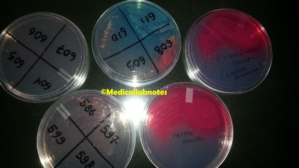
Proteus growth on CLED medium

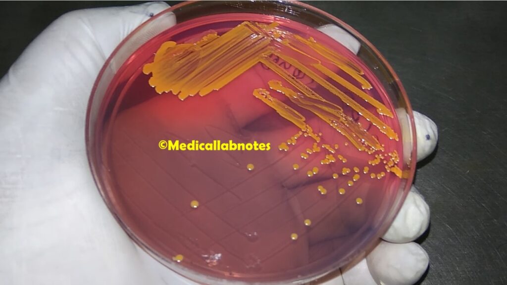
Staphylococcus aureus on CLED agar
Klebsiella pneumoniae, Pseudomonas aeruginosa, and Enterococcus colony morphology on CLED agar

E. coli, Pseudomonas aeruginosa, and Staphylococcus aureus colony characteristics on CLED agar

Other than Urine except for blood specimens
Heavy growth of Staphylococcus aureus isolated.
Light growth of Staphylococcus aureus isolated.
Moderate growth of Methicillin-resistant Staphylococcus aureus (MRSA) isolated.
Heavy growth of Escherichia coli isolated.
Heavy growth of Klebsiella pneumoniae isolated.
Heavy growth of Enterococcus species isolated.
Heavy growth of Enterococcus species isolated.
Light growth of Pseudomonas aeruginosa isolated.
Heavy growth of Pseudomonas aeruginosa isolated
Moderate growth of Proteus vulgaris isolated.
Blood Culture (no need for grading)
Salmonella Typhi/ Salmonella enterica serotype Typhi isolated.
Klebsiella pneumoniae isolated.
Salmonella Typhi isolated from blood culture

Salmonella Paratyphi isolated from blood culture

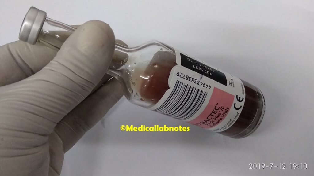
BD Bactec Blood Culture Vial
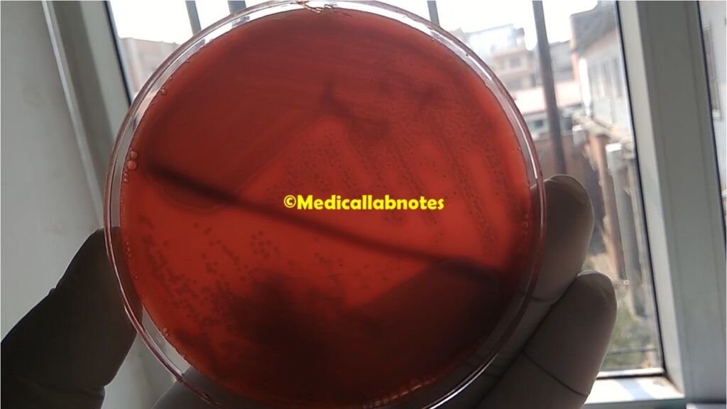
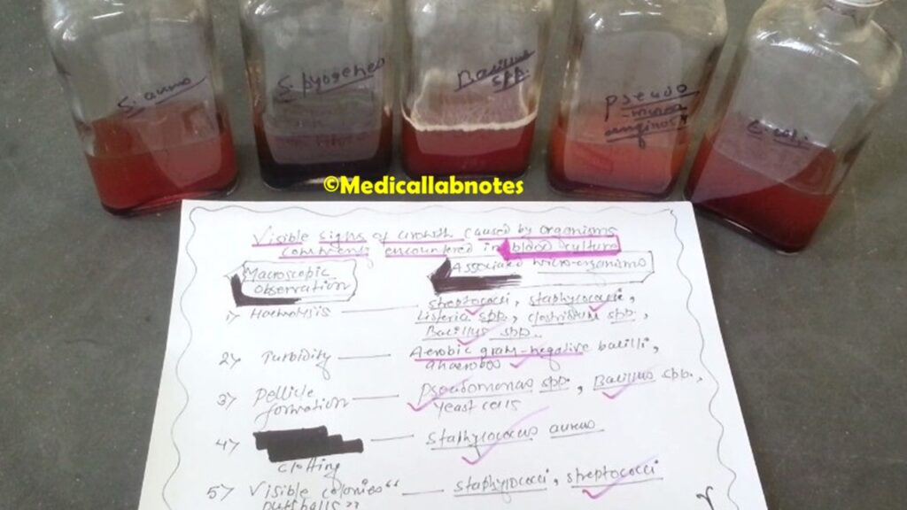
Various Bacterial Growth on Blood Culture Bottles
Keynotes on Microbiology Reporting Methods
- Urine-Contamination, please repeat the mid-stream urine (MSU) sample.
- A non-Ideal smear of Sputum-Sputum for Gram Stain
Pus cells<25/LPF
Epithelial cells>10/LPF
Organism: Normal Upper Respiratory Tract Flora Seen

- Ideal smear-Sputum for Gram Stain
Pus cells>25/LPF
Epithelial cells<10/LPF
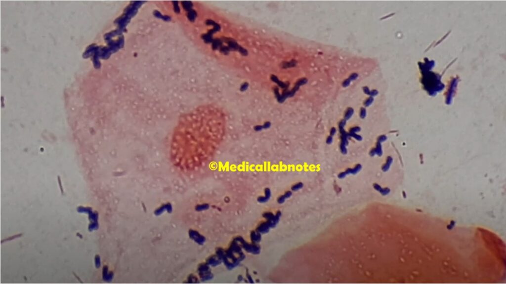
Organism: Numerous Gram-negative bacilli seen

- Body fluids-Gram stain
Pus cell: not seen
Organism: Not seen
- In the case of reporting blood culture and sensitivity- No growth after 48 hours of incubation at 37°C. There should be template notes like-Further incubation will be up to 3 days. If there is any pathogen detected, the patient or his/her visitor will be contacted for the final report.
- The remaining templates can be added according to facing the situation.
Thanks so much for the blog article.Thanks Again. Fantastic.
Thank you for sharing indeed great looking !
Nice i really enjoyed reading your blogs. Keep on posting. Thanks