
Introduction of Staphylococcus aureus
Table of Contents
Scientific classification of Staphylococcus aureus is given as:
| Domain | Bacteria |
| Phylum | Firmicutes |
| Class | Bacilli |
| Order | Bacillales |
| Family | Staphylococceae |
| Genus | Staphylococcus |
| Species | aureus |
Staphyle means bunch and Kokko mean berry. Staphylococcus aureus was isolated by Pasteur (1880) from a pus sample. The pathogenic role of Staphylococcus was described by Sir Alexander Ogston and who was a surgeon from Scotland.
Other than Staphylococcus cluster-forming cocci are-
- Micrococcus
- Gafkey
- Sarcina
- Peptococcus
- Aerococcus
Definition of Staphylococcus aureus
Spherical, non-motile, gram-positive, cluster forming. On nutrient agar, growth is opaque and golden yellow or white in color. Catalase test positive, coagulase test positive, oxidase negative, aerobic or facultative anaerobe. It is a parasite of humans and animals.
Habitat of S. aureus
Normal flora of the skin, upper respiratory tract, and feces of humans, animals, and birds too.
Morphology of Staphylococcus
- Round or spherical arranged in clusters.
- 0.8-1.0 μm in size
- Non-motile, non-sporing, usually non encapsulate while some strains are encapsulated. The capsule is of two types-
- Microcapsule <200 nm
- Macro capsule >200 nm and is responsible for the slime layer
Cultural characteristics of S. aureus
On nutrient agar
- Smooth, circular, often yellow-pigmented colonies and non-diffusible.
- 1-2 mm in diameter
- Butyrous inconsistency
On blood agar: Beta hemolytic
Pigmentation: Golden yellow and increased in the presence of CO2 and also at room temperature. Pigmentation can be induced by culturing bacteria into 30% milk agar, potato, and 1% glycerol monoacetate or phosphate agar.
Selective media for Staphylococcus
- 7-10% salt agar
- Mannitol salt agar
- Tellurite glycine agar
- Phenolphthalein phosphate agar
- Polymyxin B agar (75 μg/ml)
Resistance
The thermal death point of Staphylococcus aureus is 60°C for 30 minutes. It can survive in dried pus for 2-3 months.
Cell wall
Protein -A: It has a specific affinity for the Fc portion of the IgG molecule (except Ig3) leaving the Fab region free to combine with its specific antigen resulting in agglutination known as co-agglutination. The peptidoglycan of the cell activates complement and induces the release of inflammatory cytokines. Similarly teichoic acid of the cell wall facilitates the adhesion of the cocci to the host cell surface.
Phage types
With the use of 28 phages, several hundred phage types have been identified among them important phage types are-
- Group 1: 8052A/79 (Hospital strains)
- Group 2: 3B/3C/55 (Impetigo / Staphylococcal Scalded Syndrome)
- Group 3: 6/47 (Enterotoxin producer)
Serotype
There are 30 serotypes based on protein A antigen.
Enzyme and Toxins
Toxins
- Haemolysin: alpha, beta, gamma
- Leucodin
- Enterotoxin A-F
- Type A and B are responsible for food poisoning.
- 25 µg of toxin B can cause food poisoning.
- Epidermolysin toxin: It is responsible for Staphylococcal Scalded Syndrome (SSS) or Ritter’s disease.
- Toxic Shock Syndrome Toxin (TSST): Type -F
Enzymes
- Coagulase: It is of two types bound and free coagulase.
- Phosphatase
- DNAse
- Staphylokinase
- Hyaluronidase
- Lipase
- Protease
Pathogenicity of Staphylococcus aureus
Staphylococcus aureus can cause the following diseases-
- Abscess
- Conjunctivitis
- Corneal ulcer
- Septicemia
- Endocarditis
- Pneumonia
- Mastitis: It is an inflammation of the breast.
- Empyema: It is an accumulation of pus in the body cavity.
- Food poisoning
- Staphylococcal Scalded Syndrome
- Toxic Shock Syndrome (TSS)-enterotoxin F
- Septic arthritis
- Meningitis
- Osteomyelitis
Laboratory Diagnosis of Staphylococcus aureus
Samples/specimens collection: It depends on the site of infection and the nature of the lesion. e.g.
Pus (Suppurative lesion)
CSF ( meningitis)
Blood (septicemia)
Sputum( respiratory infection)
Nasal swab (detection of carriers)
Feces and remains of food (food poisoning)
Gram stain: Gram-positive cocci in singles, pairs, and clusters.
Culture
Media -for routine Nutrient agar and blood agar
for Selective
- 7-10% salt agar
- Mannitol salt agar
- Tellurite glycine agar
- Phenolphthalein phosphate agar
- Polymyxin B agar (75 μg/ml)
Colony characteristics
- Smooth, circular, often yellow-pigmented colonies and non-diffusible.
- 1-2 mm in diameter
- Butyrous inconsistency
Beta hemolytic colony
Biochemical tests of S. aureus
Catalase test: Positive
Oxidation and fermentation (OF) test: Fermentative
Coagulase test: Positive
DNAse test: Positive
From these features, the organism is identified as Staphylococcus aureus.
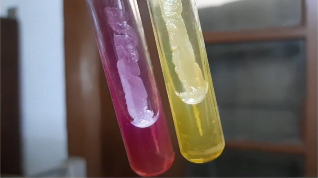
Coagulase test
Coagulase brings about the clotting of plasma which is similar to the thrombin-catalytic conversion of fibrinogen into fibrin.
Types
- Free coagulase: This is an extracellular enzyme of bacteria secreted into the medium. It is a thrombin-like substance that can change fibrinogen to fibrin. A tube coagulase test is performed for its detection.
- Bound coagulase: This is closely bound to the cell wall. On its surface, it has receptors for fibrinogen so fibrin forms links between the bacteria. This causes the clumping of Staphylococci. Hence, bound coagulase is also known as the clumping factor. A slide coagulase test is done for its detection.
Treatment
Following antibiotics are available for antibiotics sensitivity testing (AST)-
- Clindamycin
- Erythromycin
- Cefoxitin
- Chloramphenicol
- Ciprofloxacin
- Gentamycin
- Ofloxacin
- Cotrimoxazole
- Doxycycline
- Vancomycin
- Teicoplanin
- Linezolid
- Nitrofurantoin
Keynotes on Staphylococcus
- Nitrofurantoin is only applicable in case of urinary tract infection replacing chloramphenicol.
- To treat Methicillin-resistant Staphylococcus aureus (MRSA), vancomycin is recommended whereas to treat Vancomycin-resistant Staphylococcus aureus (VRSA), linezolid is preferred.
Staphylococcus Pertaining Footages
Gram-positive cocci in singles, pairs, and clusters of Staphylococcus in Gram-staining of clinical sample

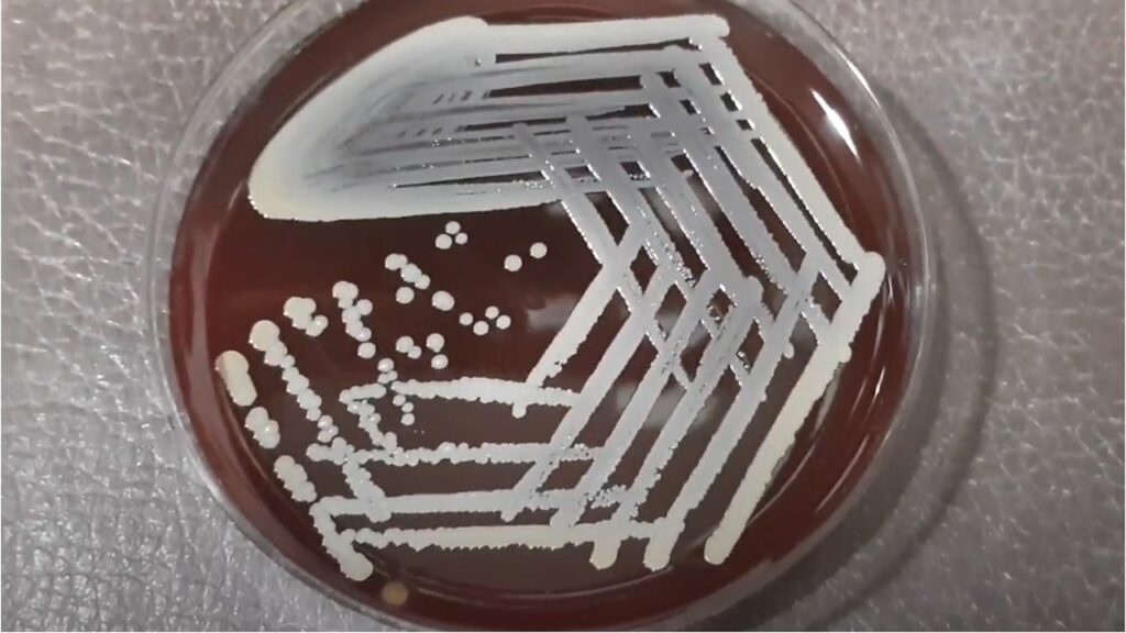
Staphylococcus aureus on blood agar
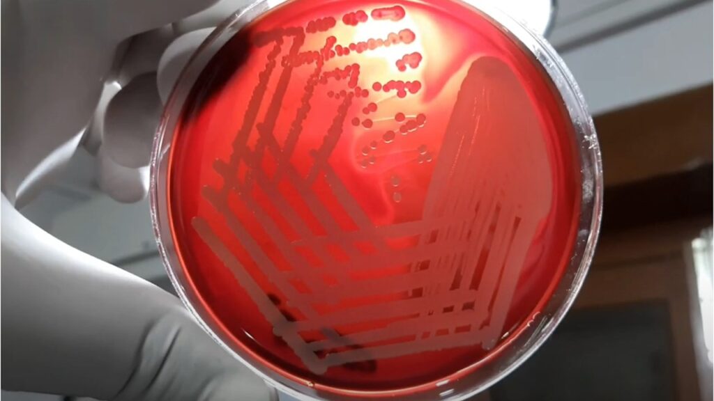
Beta-haemolytic colonies of Staphylococcus aureus on blood agar
GPC in singles, pairs, chains, and clusters of Staphylococcus aureus in Gram staining of culture

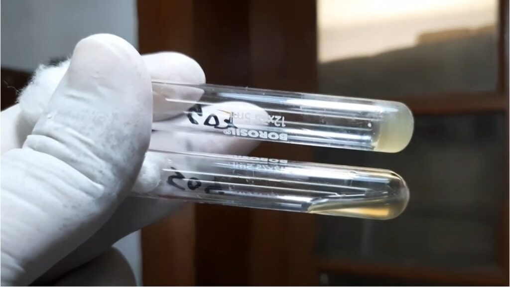
Coagulase test postive of Staphylococcus aureus
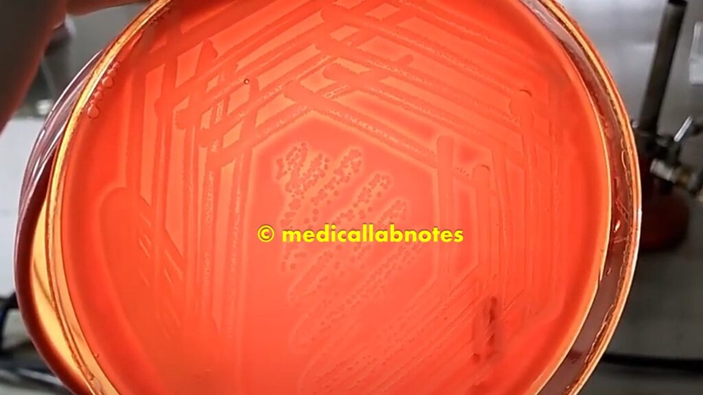
Staphylococcus aureus (yellow) and Coagulase Negative Staphylococci (CoNS) growth on Mannitol Salt Agar (MSA)
Further Readings
- Bailey & Scott’s Diagnostic Microbiology. Editors: Bettey A. Forbes, Daniel F. Sahm & Alice S. Weissfeld, 12th ed 2007, Publisher Elsevier.
- Colour Atlas and Textbook of Diagnostic Microbiology. Editors: Koneman E.W., Allen D.D., Dowell V.R. Jr, and Sommers H.M.
- Clinical Microbiology Procedure Handbook, Chief in editor H.D. Isenberg, Albert Einstein College of Medicine, New York, Publisher ASM (American Society for Microbiology), Washington DC.
- Jawetz, Melnick and Adelberg’s Medical Microbiology. Editors: Geo. F. Brook, Janet S. Butel & Stephen A. Morse, 21st ed 1998, Publisher Appleton & Lance, Co Stamford Connecticut.
- Manual of Clinical Microbiology. Editors: P.R. Murray, E. J. Baron, M. A. Pfaller, F. C. Tenover, and R. H. Yolken, 7th ed 2005, Publisher ASM, USA
- Mackie and Mc Cartney Practical Medical Microbiology. Editors: J.G. Colle, A.G. Fraser, B.P. Marmion, A. Simmons, 4th ed, Publisher Churchill Living Stone, New York, Melbourne, Sans Francisco 1996.
- Textbook of Diagnostic Microbiology. Editors: Connie R. Mahon, Donald G. Lehman & George Manuselis, 3rd edition2007, Publisher Elsevier.
- District Laboratory Practice in Tropical Countries – Part-2- Monica Cheesebrough- 2nd Edn Update
- Topley & Wilsons’ Principle of Bacteriology, Virology, and immunology. Editors: M.T. Parker & L.H. Collier, 8th ed 1990, Publisher Edward Arnold publication, London.