
Introduction
Table of Contents
The Dalmau plate technique is a classic mycological method used to identify and differentiate yeast species, specifically Candida species, by observing their unique morphological features.

The technique is a specialized culture method that uses a “starvation medium” to induce the formation of characteristic structures like pseudohyphae, blastoconidia, and chlamydospores. It remains a valuable, cost-effective tool for preliminary identification in clinical laboratories.
Principle
The principle relies on using a nutrient-deficient medium (starvation medium) and creating a microaerophilic environment (reduced oxygen) by placing a coverslip over the inoculated area. This stress induces yeasts to switch from their vegetative budding state to filamentous growth or spore formation.

Test Requirements
- Media: Cornmeal Agar (CMA) is most common; Rice extract agar or Pea agar can also be used.
- Additive: Tween 80 (a surfactant) is often added to reduce surface tension and enhance the formation of pseudohyphae and chlamydospores.
- Equipment: Sterile inoculation wire or loop, sterile glass coverslips, and a light microscope.

Procedure
- Prepare Cornmeal agar containing 1% Tween 80 in a 90-mm plate.
- Divide the plate into 4 quadrants and label each quadrant.
- Using a sterile needle or straight wire, lightly touch the yeast colony and then make 2-3 streaks of approximately 3.5 – 4 cm long and 1.2 cm apart.
- Place a flame-sterilized and cooled 22 mm square cover glass over the control part of the streak.
- This will provide a partially anaerobic environment at the margins of the cover slip. Incubate the plates at 25°C for 3-5 days.
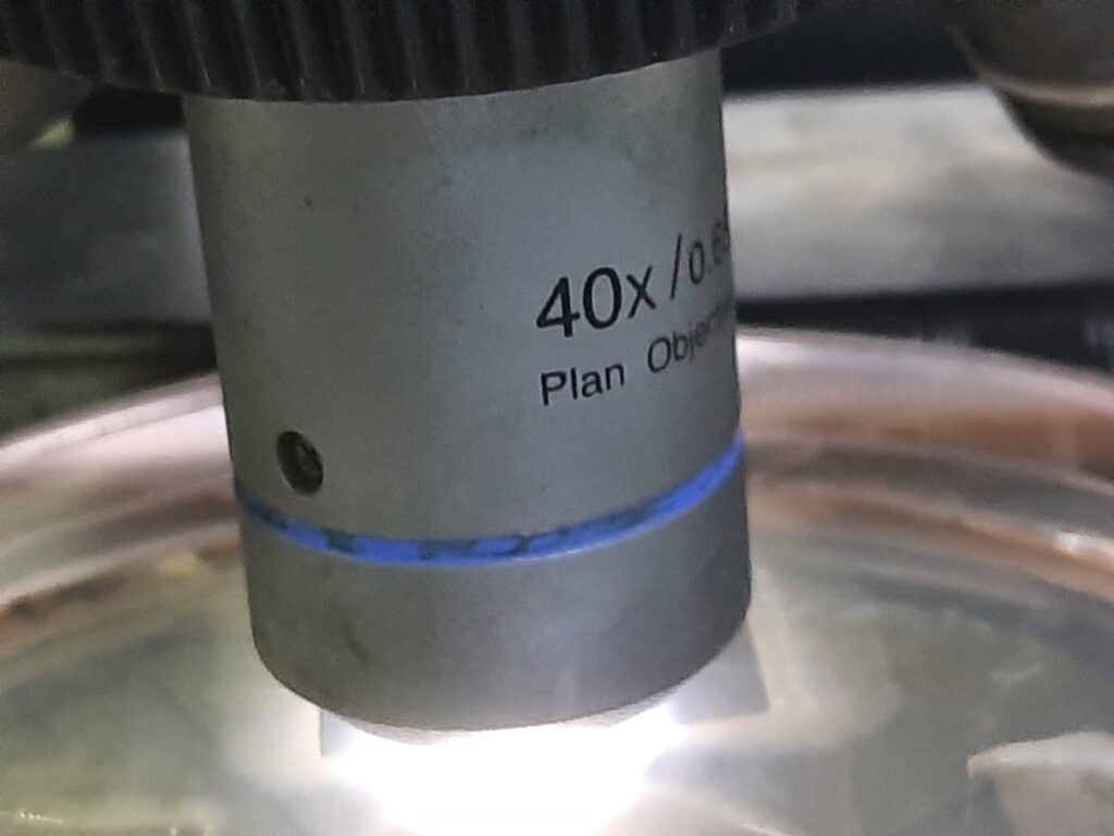
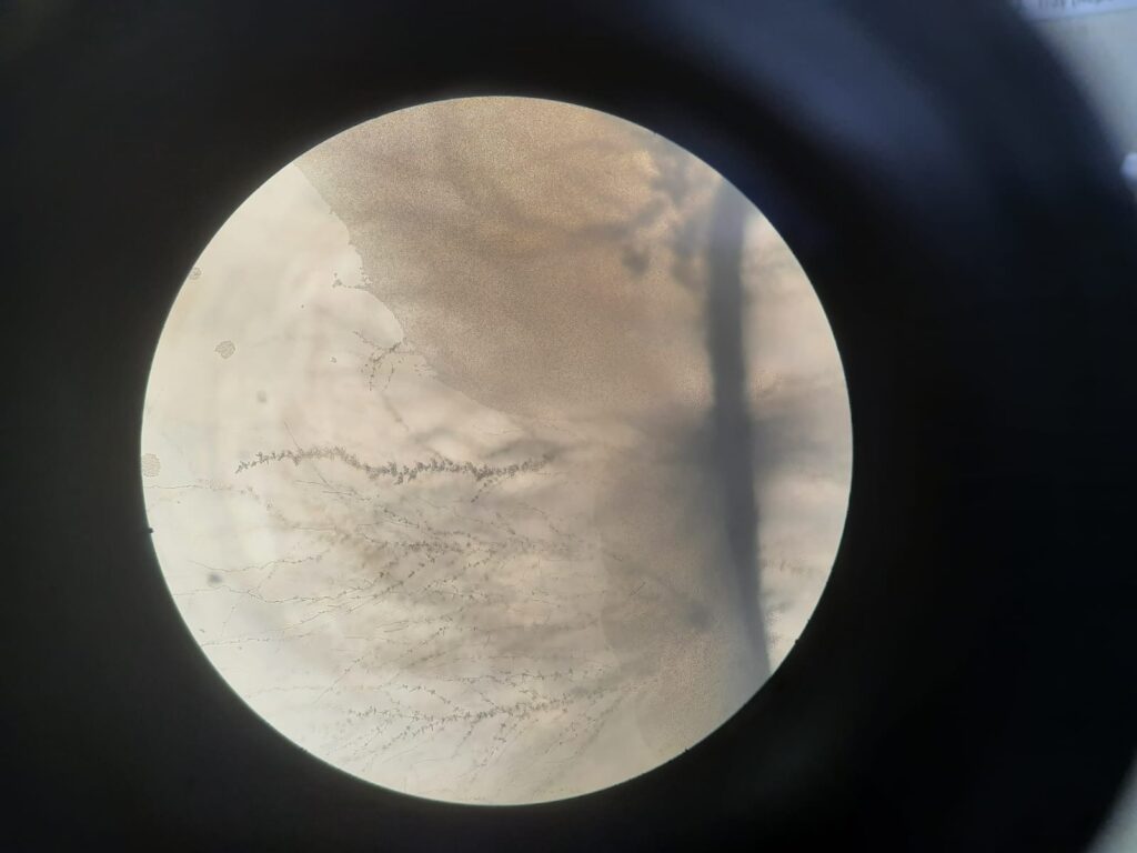
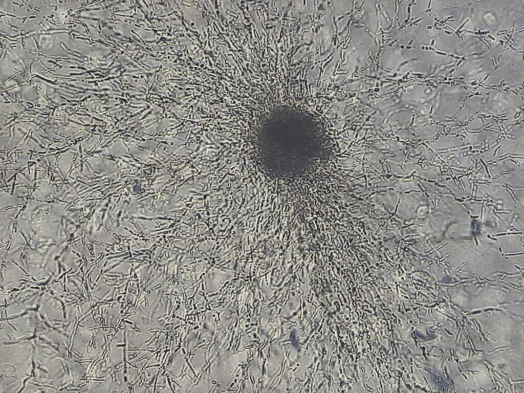
- Remove the lid of the petri plate and place the plate on the microscope stage, and observe the edge of the cover glass using the low-power objective (10X) first and then the high-power objective (40X).
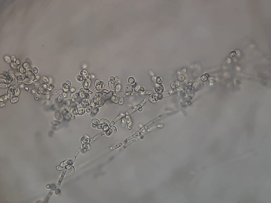
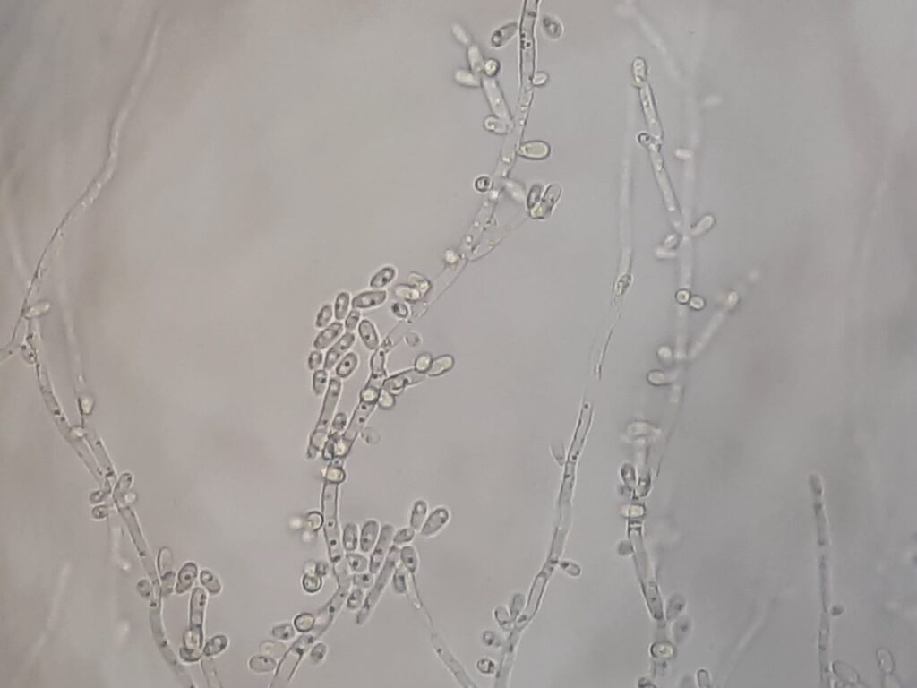
- Note morphological features like hyphae, pseudohyphae, blastospores, ascospores, chlamydospores, basidiospores, or sporangia, and find out the organism as shown in the application table.
Applications
- Speciation of Candida: Differentiating C. albicans (which produces large, thick-walled terminal chlamydospores) from non-albicans species.
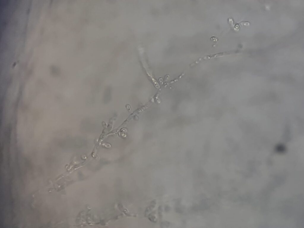
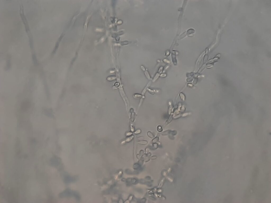
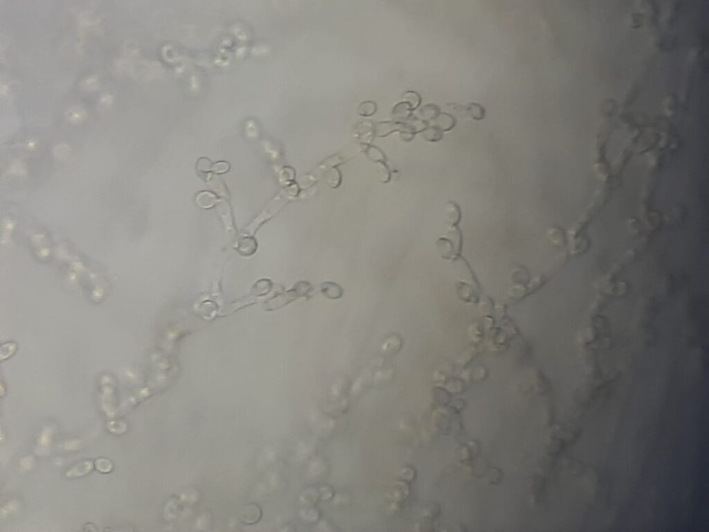
- Morphological Studies: Observing the arrangement of blastoconidia along pseudohyphae, which is unique to different yeast genera.
- Starvation Response: Studying how yeast-like fungi adapt to nutrient-poor environments.
| Feature | Possibility |
| Terminal chlamydoconidia | Candida albicans, Candida dubliniensis |
| Yeasts only | Candida glabrata, Candida famata, Pichia anomala, Pichia augusta (Hansenula polymorpha), Cryptococcus neoformans |
| Giant hyphae, blastospores at nodes | Candida parapsilosis |
| Abundant pseudohyphae, pine forest arrangement, blastoconidia formed at or in between septa | Candida tropicalis |
| Elongated yeasts, abundant pseudohyphae (matchstick-like appearance) | Candida krusei (Pichia kudriavzevii) |
| Scant pseudohyphae with chains of blastoconidia | Candida guilliermondii |
| Short, distinctly curved pseudohyphae with occasional blastoconidia at septa | Candida lusitaniae |
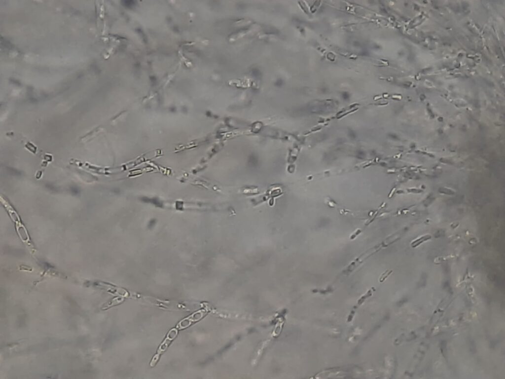
| Arthroconidia without blastoconidia | Geotrichum species |
| Arthroconidia with blastoconidia | Trichosporon species |


Keynotes
- Observation Point: The most diagnostic morphology is typically found at the edge of the coverslip, where the oxygen gradient is ideal.
- Chlamydospores: Candida dubliniensis often produces multiple terminal chlamydospores in clusters, while Candida albicans typically produces them singly.
- Alternative Methods: While reliable, it is slower than modern methods like the Germ Tube Test or MALDI-TOF MS.
Further Readings
- https://www.microdigest.net/2025/09/dalmau-plate-technique-cherished-in.html
- https://mycology.adelaide.edu.au/fungal-descriptions-and-antifungal-susceptibility/yeast-like-fungi
- https://www.afwgonline.com/wp-content/uploads/2018/12/1115_TanAL_Identification-of-Yeasts-lecture_Final.pdf
- https://mycology.adelaide.edu.au/candida
- https://www.remedypublications.com/open-access/identification-of-candida-species-conventional-methods-in-the-era-of-molecular-diagnosis-775.pdf
- https://www.sciencedirect.com/topics/pharmacology-toxicology-and-pharmaceutical-science/chlamydospore
- https://www.sciencedirect.com/science/article/pii/S0732889323001281
- https://mycology.adelaide.edu.au/fungal-descriptions-and-antifungal-susceptibility/yeast-like-fungi/cryptococcus
- https://www.researchgate.net/figure/The-characteristic-Dalmau-morphology-in-different-Candida-spp_tbl1_342505552
- https://pmc.ncbi.nlm.nih.gov/articles/PMC11210618/
- https://www.icmr.gov.in/icmrobject/custom_data/pdf/resource-guidelines/Mycology_SOP_2nd_Ed_2019.pdf
- https://www.sciencedirect.com/topics/biochemistry-genetics-and-molecular-biology/methylotrophic-yeast