
Introduction
Table of Contents
Pseudomonas aeruginosa is a Gram-negative bacterium that is widely distributed in nature and is known for its versatility and adaptability. It is an opportunistic pathogen capable of causing infections in humans, particularly in individuals with weakened immune systems or underlying medical conditions. P. aeruginosa is often associated with nosocomial (hospital-acquired) infections and is a leading cause of morbidity and mortality in healthcare settings.

One of the distinguishing characteristics of P. aeruginosa is its ability to survive in diverse environments, including soil, water, and hospital settings. This bacterium has a remarkable arsenal of virulence factors, such as toxins, enzymes, and adhesins, which enable it to establish infections and evade the immune system.
P. aeruginosa infections can manifest in various forms, including respiratory tract infections (such as pneumonia), urinary tract infections, skin and soft tissue infections, and bloodstream infections. It is also a common cause of chronic infections, particularly in individuals with cystic fibrosis.
Due to its intrinsic resistance to many antibiotics and its ability to acquire additional resistance mechanisms, P. aeruginosa infections can be challenging to treat. This bacterium produces various enzymes that can degrade antibiotics, and it can also form biofilms, which are complex communities of bacteria embedded in a protective matrix. Biofilms enhance the bacterium’s resistance to antibiotics and host defenses, further complicating treatment.

Prevention and control of P. aeruginosa infections rely on strict infection control measures, including proper hand hygiene, disinfection of equipment, and surveillance of healthcare settings. In addition, antibiotic stewardship programs aim to minimize the overuse and misuse of antibiotics, which can contribute to the development of drug-resistant strains.
In summary, Pseudomonas aeruginosa is a versatile opportunistic pathogen that can cause a range of infections, especially in healthcare settings. Its ability to survive in diverse environments, its arsenal of virulence factors, and its resistance to antibiotics make it a significant clinical concern. Understanding its biology and implementing appropriate preventive measures are crucial for managing and controlling P. aeruginosa infections.
Morphology
Pseudomonas aeruginosa is a Gram-negative bacterium that exhibits a distinct morphology. Here is an overview of its morphology:
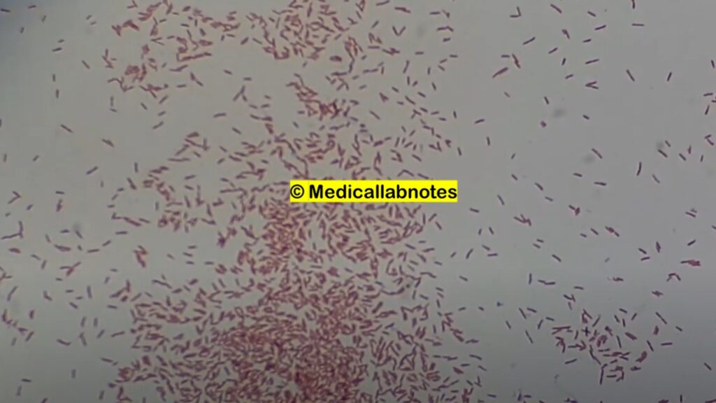
- Shape: It is a rod-shaped bacterium, commonly referred to as a bacillus. The cells are elongated and have a cylindrical or slightly curved appearance.

- Size: The size of its cells can vary, but they are typically around 1 to 3 micrometers in length and 0.5 to 1 micrometer in width.
- Arrangement: Its cells usually occur as single, non-motile rods. They can also form short chains or clusters in certain conditions.
- Flagella: P. aeruginosa is motile, possessing one or more polar flagella. These flagella allow the bacterium to move and explore its environment.
- Capsule: P. aeruginosa is known to produce a polysaccharide capsule, which surrounds the bacterial cell. This capsule aids in protecting the bacterium from host immune responses and contributes to its virulence.
- Pigmentation: P. aeruginosa colonies often display various pigments, ranging from green to yellow or blue-green. These pigments, such as pyocyanin and pyoverdine, are secondary metabolites produced by the bacterium and can contribute to its characteristic coloration.

- Biofilm formation: P. aeruginosa has a remarkable ability to form biofilms. In this mode, the bacteria aggregate and adhere to surfaces, forming complex communities encased in a self-produced extracellular matrix. Biofilm formation is facilitated by the production of extracellular polysaccharides and contributes to the bacterium’s resistance to antibiotics and host defenses.
Pathogenicity
Pseudomonas aeruginosa is a highly adaptable and opportunistic pathogen that is capable of causing a wide range of infections in humans. Its pathogenicity is attributed to a combination of factors, including its ability to produce a variety of virulence factors and its intrinsic and acquired antibiotic resistance mechanisms. Here are some key aspects of the pathogenicity of P. aeruginosa:
- Virulence Factors: It possesses an extensive arsenal of virulence factors that enable it to establish and maintain infections. These include:a. Exotoxins: It produces several exotoxins, such as exotoxin A and exoenzymes S, T, and U. These toxins interfere with host cell functions, disrupt cellular signaling pathways, and contribute to tissue damage and inflammation.b. Enzymes: It produces enzymes, including proteases, elastases, and phospholipases, which degrade host tissues and extracellular matrix components, promoting tissue invasion and immune evasion.c. Pyocyanin: Pyocyanin is a blue-green pigment produced by P. aeruginosa. It has toxic effects on host cells, contributes to oxidative stress, and impairs immune cell function.d. Alginate: P. aeruginosa can produce alginate, a polysaccharide that forms the extracellular matrix of biofilms. Biofilms protect the bacterium from immune responses and antimicrobial agents.
- Antibiotic Resistance: It is intrinsically resistant to many antibiotics due to its impermeable outer membrane, efflux pumps that expel drugs, and enzymes that can degrade antibiotics. Furthermore, it has a remarkable ability to acquire additional resistance through the acquisition of resistance genes, making treatment challenging.
- Biofilm Formation: It is known for its ability to form biofilms on both biotic and abiotic surfaces. Biofilms enhance bacterial survival, as they provide protection against host immune defenses and antibiotics. In the context of infections, biofilms can form on medical devices, such as catheters or ventilator tubes, leading to persistent and difficult-to-treat infections.
- Host Immune Response: It can evade and manipulate host immune responses. It produces factors that interfere with phagocytosis, suppress immune cell activity, and modulate host immune signaling pathways, allowing the bacterium to persist and cause chronic infections.
The pathogenicity of P. aeruginosa is influenced by the interplay of these virulence factors, antibiotic resistance mechanisms, and the host’s immune status. In individuals with compromised immune systems, such as those with cystic fibrosis, burn wounds, or immunosuppression, P. aeruginosa infections can be particularly severe and difficult to treat. Effective management of P. aeruginosa infections involves a combination of targeted antibiotic therapy, removal of infected devices, and infection control measures to prevent the spread of the bacterium.
Lab Diagnosis
The laboratory diagnosis of Pseudomonas aeruginosa infections involves several methods aimed at identifying the bacterium and determining its antibiotic susceptibility. Here are some commonly used techniques:
- Gram Staining: It appears as Gram-negative rods under a microscope. Gram staining is a rapid initial step that helps differentiate Pseudomonas species from other bacteria based on their cell wall characteristics.
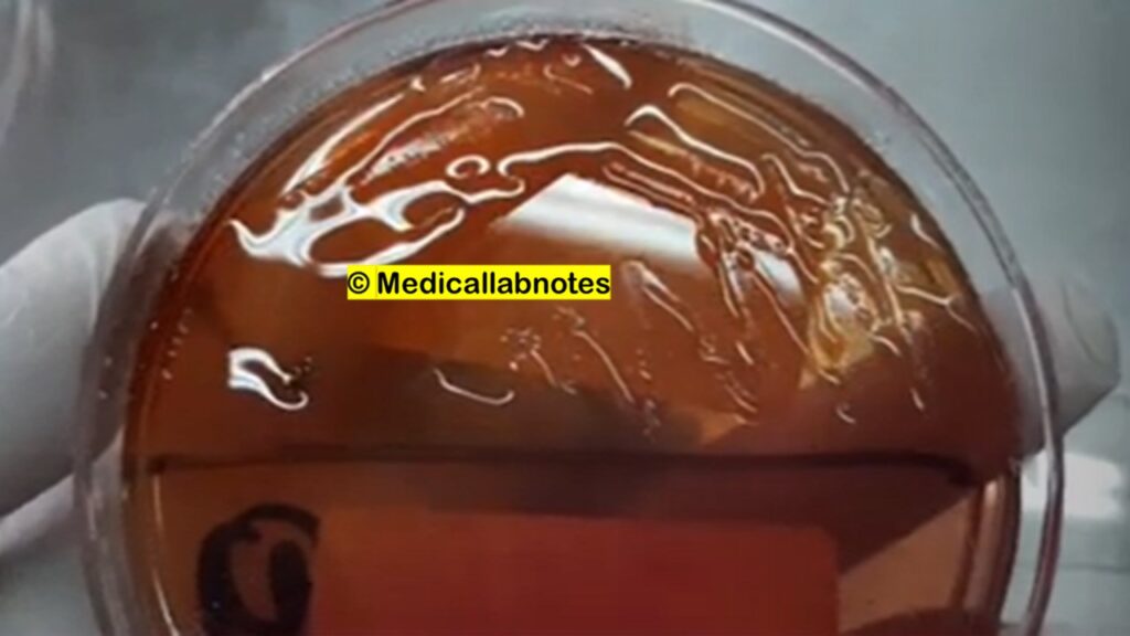
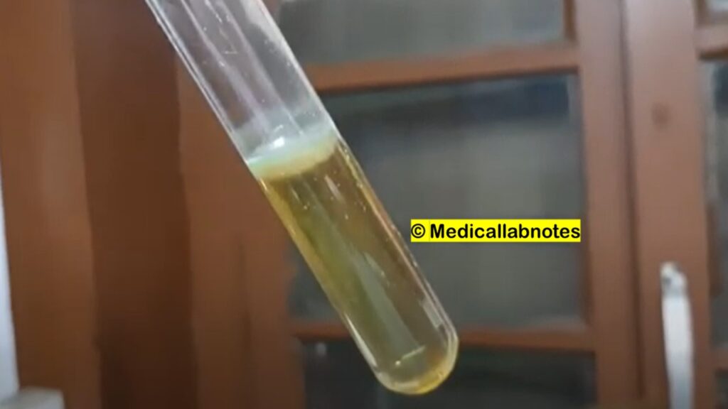
- Culture and Isolation: The primary method for diagnosing P. aeruginosa is by culturing the bacterium from clinical specimens. The specimen may include sputum, urine, wound swabs, blood, or other relevant samples. The specimen is streaked onto selective media such as MacConkey agar or cetrimide agar, which promote the growth of Pseudomonas species. It typically exhibits distinctive characteristics on culture, including a characteristic odor (described as grape-like or fruity) and blue-green pigment production.

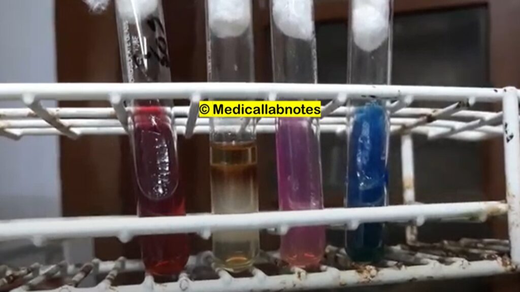
- Biochemical Tests: Various biochemical tests are performed to confirm the identity of P. aeruginosa. These tests include oxidase positivity (P. aeruginosa is oxidase-positive), catalase positivity, and the ability to utilize specific carbon sources, such as glucose and lactose.
- Microscopy: Microscopic examination of the cultured bacteria can provide additional information. Its cells can be observed as motile rods with polar flagella using techniques like phase-contrast microscopy or dark-field microscopy.
- Identification Systems: Commercial identification systems, such as the API system or Vitek system, can be employed to confirm the identity of P. aeruginosa. These systems utilize a panel of tests and biochemical reactions to identify the bacterium based on its unique metabolic profile.
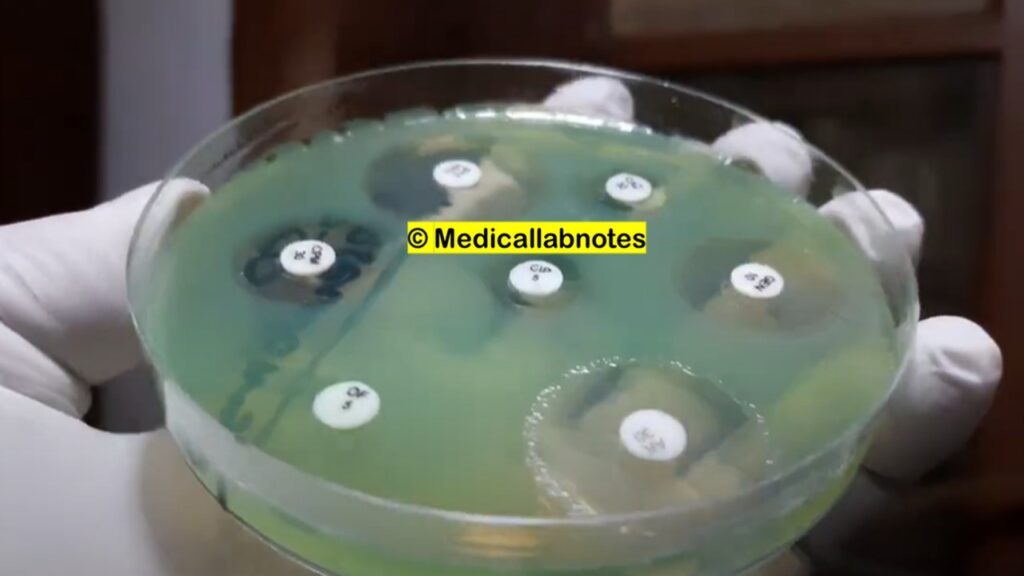
- Antibiotic Susceptibility Testing: P. aeruginosa‘s intrinsic and acquired antibiotic resistance mechanisms make it important to determine its susceptibility to antimicrobial agents. This is typically done through methods like disk diffusion or broth microdilution. The results guide the selection of appropriate antibiotics for treatment.
Treatment
The treatment of Pseudomonas aeruginosa infections can be challenging due to the bacterium’s intrinsic and acquired antibiotic resistance mechanisms. The choice of treatment depends on several factors, including the site and severity of infection, the patient’s overall health, and the antibiotic susceptibility profile of the specific P. aeruginosa strain. Here are some commonly used approaches:
- Antibiotic Therapy: P. aeruginosa infections are typically treated with antibiotics. However, due to the high prevalence of resistance, susceptibility testing should be performed to guide appropriate antibiotic selection. Antimicrobial agents commonly used against P. aeruginosa include:a. Beta-lactam antibiotics: Antipseudomonal penicillins (such as piperacillin-tazobactam) and cephalosporins (such as ceftazidime or cefepime) are often used as first-line agents. However, resistance to these drugs has been observed, so susceptibility testing is crucial.b. Carbapenems: Drugs like imipenem, meropenem, or doripenem are effective against P. aeruginosa and are often used when other beta-lactams are ineffective.c. Fluoroquinolones: Ciprofloxacin and levofloxacin are fluoroquinolones with activity against P. aeruginosa. However, resistance can develop rapidly, so they are often used in combination with other agents.d. Aminoglycosides: Agents such as gentamicin or tobramycin may be used in combination with beta-lactams or carbapenems for synergistic effects.e. Polymyxins: Colistin (polymyxin E) and polymyxin B are considered last-resort options for multidrug-resistant P. aeruginosa infections. However, their use is limited due to associated toxicity and the emergence of resistance.It’s important to note that combination therapy with two or more antibiotics is often employed for severe P. aeruginosa infections, especially in critically ill patients or those with high-risk factors.
- Removal of Infected Devices: In cases where P. aeruginosa infection is associated with medical devices such as catheters or ventilator tubes, removal or replacement of the infected device is essential for successful treatment.
- Infection Control Measures: Strict adherence to infection control practices is crucial to prevent the spread of P. aeruginosa infections, particularly in healthcare settings. This includes proper hand hygiene, disinfection of equipment, and adherence to appropriate infection control protocols.
- Adjunctive Therapies: In some cases, adjunctive therapies may be employed to support the treatment of P. aeruginosa infections. These may include wound care, surgical intervention to remove infected tissues, or therapies targeting specific complications (e.g., respiratory support for pneumonia).

Prevention
Prevention of Pseudomonas aeruginosa infections involves a combination of strategies to reduce the risk of exposure and transmission. Here are some key preventive measures:
- Infection Control Practices in Healthcare Settings:
- Hand Hygiene: Strict adherence to hand hygiene protocols, including handwashing with soap and water or using alcohol-based hand sanitizers, is crucial in preventing the transmission of P. aeruginosa. Healthcare workers should perform hand hygiene before and after patient contact and follow proper hand hygiene techniques.
- Personal Protective Equipment (PPE): Healthcare workers should use appropriate PPE, such as gloves, gowns, masks, and eye protection, when providing care to patients with known or suspected P. aeruginosa infections.
- Environmental Cleaning: Regular and effective cleaning and disinfection of surfaces and equipment in healthcare settings help prevent the transmission of P. aeruginosa. Use of appropriate disinfectants and following recommended cleaning protocols is essential.
- Catheter and Device Care:
- Central Venous Catheters (CVCs): Strict adherence to sterile insertion techniques, proper catheter site care, and regular assessment of catheter necessity are important in preventing P. aeruginosa bloodstream infections associated with CVCs.
- Urinary Catheters: Proper insertion and maintenance of urinary catheters, along with regular assessment and prompt removal when no longer necessary, help minimize the risk of urinary tract infections caused by P. aeruginosa.
- Ventilator Care: Proper care and maintenance of ventilator equipment, including regular cleaning and disinfection, help prevent P. aeruginosa respiratory tract infections in ventilated patients.
- Water and Environmental Control:
- Water Systems: Ensuring proper maintenance and disinfection of water systems in healthcare facilities, including regular monitoring for P. aeruginosa colonization, is important to prevent waterborne transmission.
- Pools and Hot Tubs: Proper chlorination and maintenance of pools, hot tubs, and recreational water facilities help prevent P. aeruginosa infections associated with recreational water exposure.
- Infection Control in Specific Settings:
- Burn Units: Adherence to strict infection control measures in burn units, including hand hygiene, wound care, and aseptic techniques, is crucial to prevent P. aeruginosa infections in burn patients.
- Intensive Care Units (ICUs): Implementing comprehensive infection control practices, including surveillance, isolation precautions, and antibiotic stewardship, is important in preventing the spread of P. aeruginosa infections in ICUs.
- Antibiotic Stewardship:
- Rational use of antibiotics and adherence to antibiotic stewardship principles help minimize the development of antibiotic-resistant strains of P. aeruginosa.
It is important for healthcare facilities to have infection control protocols in place, provide training to healthcare workers on preventive measures, and maintain vigilant surveillance to promptly identify and manage P. aeruginosa infections.
Keynotes
Here are some keynotes on Pseudomonas aeruginosa:

- Pseudomonas aeruginosa is a Gram-negative bacterium known for its versatility and adaptability.
- It is an opportunistic pathogen that can cause infections in individuals with weakened immune systems or underlying medical conditions.
- P. aeruginosa is commonly associated with nosocomial (hospital-acquired) infections and is a leading cause of morbidity and mortality in healthcare settings.
- The bacterium has a wide distribution in nature and can survive in diverse environments, including soil, water, and hospital settings.
- P. aeruginosa possesses an extensive arsenal of virulence factors, such as toxins, enzymes, and adhesins, which contribute to its pathogenicity.
- It can cause a variety of infections, including respiratory tract infections, urinary tract infections, skin and soft tissue infections, and bloodstream infections.
- P. aeruginosa infections can be challenging to treat due to its intrinsic resistance to many antibiotics and its ability to acquire additional resistance mechanisms.
- The bacterium is capable of forming biofilms, which are complex communities of bacteria embedded in a protective matrix. Biofilms enhance its resistance to antibiotics and host defenses.
- Prevention and control of P. aeruginosa infections involve strict infection control measures, such as proper hand hygiene, disinfection of equipment, and surveillance in healthcare settings.
- Antibiotic stewardship programs aim to minimize the overuse and misuse of antibiotics to prevent the emergence and spread of drug-resistant P. aeruginosa strains.
Further Readings
- “Pseudomonas aeruginosa Infections: Epidemiology, Treatment, and Prevention” – A comprehensive review published in Clinical Microbiology Reviews that covers various aspects of P. aeruginosa infections, including epidemiology, pathogenesis, diagnosis, and treatment.
- “Pseudomonas aeruginosa: Resistance and Therapeutic Options at the Turn of the New Millennium” – An article published in Clinical Microbiology and Infection that discusses the antibiotic resistance mechanisms of P. aeruginosa and therapeutic options for managing infections.
- “Pseudomonas aeruginosa: Host Defence in Lung Infections” – A review article published in Respirology that focuses on the interaction between P. aeruginosa and the host immune response in respiratory tract infections, particularly in individuals with cystic fibrosis.
- “Biofilms and Antibiotic Resistance of Pseudomonas aeruginosa” – A research article published in International Journal of Antimicrobial Agents that explores the role of biofilms in P. aeruginosa infections and their impact on antibiotic resistance and treatment outcomes.
- “Pseudomonas aeruginosa: An Opportunistic Pathogen in Healthcare Settings” – A chapter in the book “Healthcare-Associated Infections” that provides an overview of P. aeruginosa infections in healthcare settings, including epidemiology, risk factors, and preventive measures.
I like this web site so much, bookmarked.
What’s up everyone, it’s my first pay a visit at this website, and post is truly fruitful in favor of me, keep up posting these articles.
Just wish to say your article is as amazing. The clarity for your publish is just
spectacular and i could think you’re knowledgeable on this subject.
Fine along with your permission let me to clutch your feed to stay updated with coming near near post.
Thank you a million and please keep up the gratifying work.