
Visual Observation
Table of Contents
- Medium: The tubes appear to be MGIT (Mycobacteriological Growth Indicator Tubes), which contain a liquid broth (Middlebrook 7H9) supplemented with OADC with antitubercular drugs, as shown in this image.
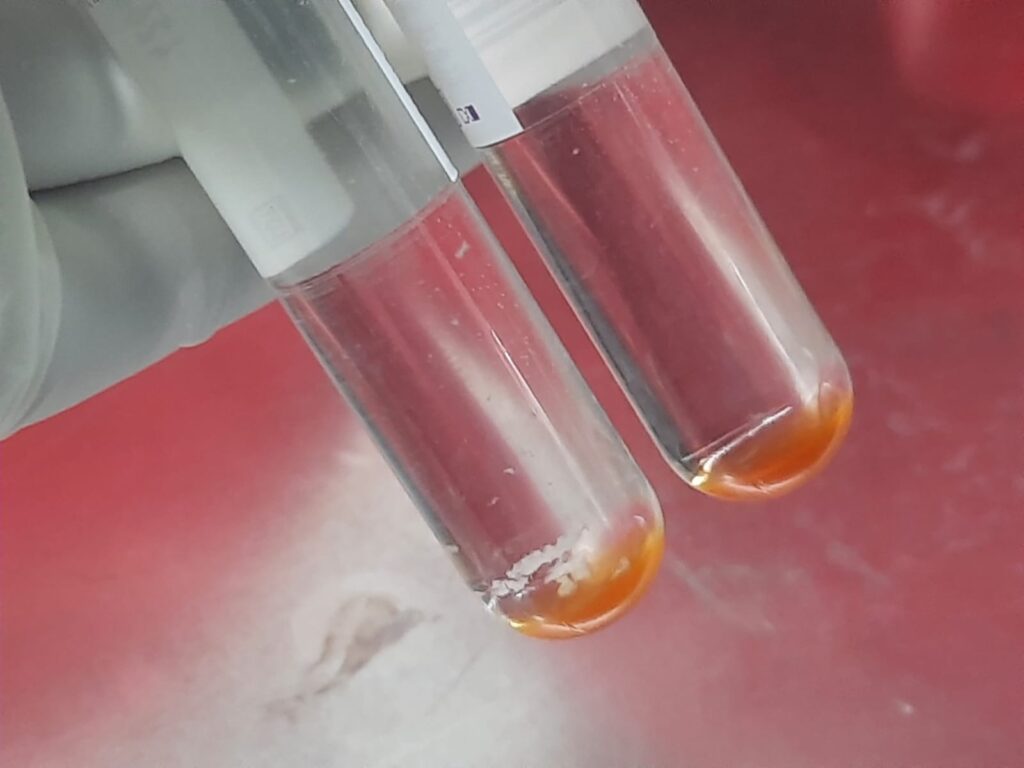
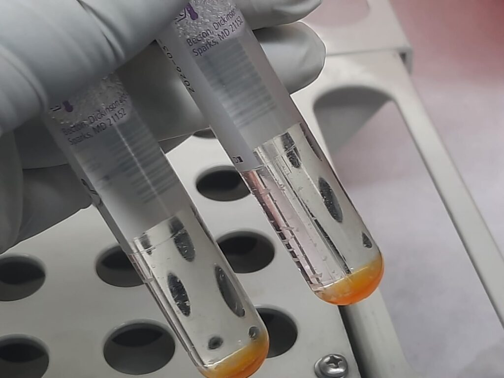
- Sediment/Growth: At the bottom of the tubes, there is a distinct orange-yellowish sediment.
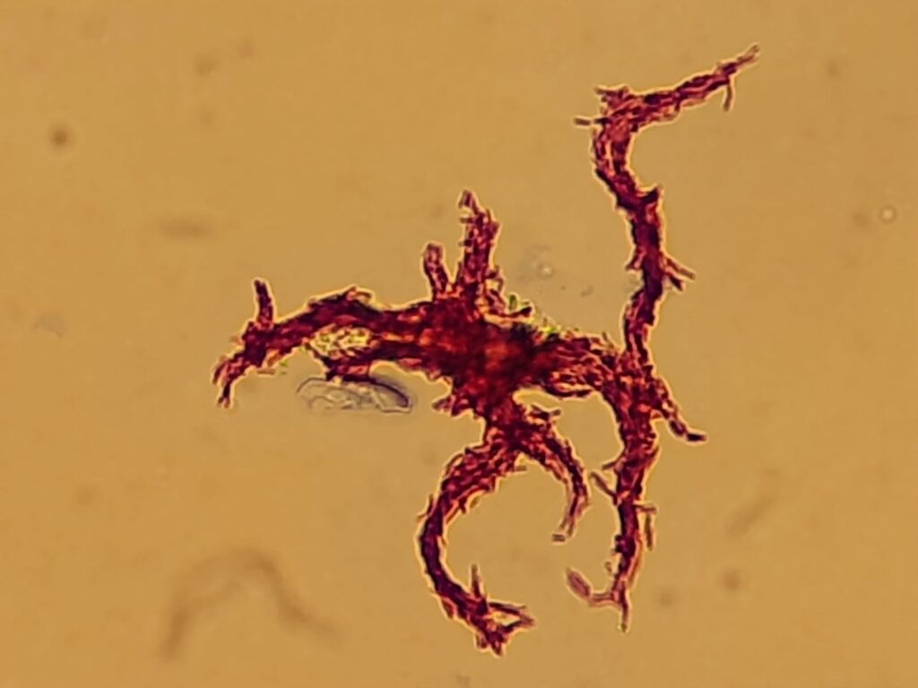
- Clumping (Cording): In the left tube, particularly, you can see white, granular, “crumb-like” aggregates or flakes. This is a classic macroscopic feature of M. tuberculosis growth in liquid media, often correlating with the “cording” phenomenon seen microscopically.
- Supernatant: The liquid above the sediment remains relatively clear, which is typical for slow-growing mycobacteria compared to the rapid, uniform turbidity seen with common bacterial contaminants.
Technical Interpretation
The MGIT Principle: These tubes contain a fluorescent sensor embedded in silicon at the bottom. As the MTBC metabolizes nutrients, it consumes dissolved oxygen.
- Initial State: High dissolved oxygen quenches the sensor’s emissions.
- Growth State: As the bacteria deplete it, the sensor begins to fluoresce under UV light.
- Result: The presence of the granular sediment you see confirms that the machine likely flagged these as “Positive.”
MTBC Characteristics
The “buff-colored” (off-white to light yellow) and granular appearance is highly indicative of the M. tuberculosis complex. Unlike Mycobacterium avium or other NTMs (Non-Tuberculous Mycobacteria), which may produce smoother or more pigmented growth, MTBC tends to form these tight, non-homogenous clumps due to the cord factor (trehalose 6,6′-dimycolate) in its cell wall.
Next Steps
Standard laboratory protocol for a positive MGIT tube usually involves:

- Ziehl-Neelsen (ZN) or Auramine-Rhodamine Staining: To confirm the presence of Acid-Fast Bacilli (AFB) and check for serpentine cording.
- Blood Agar Subculture: To rule out rapid-growing bacterial contamination.
- Rapid Immunochromatographic Test (e.g., MPT64 antigen): To differentiate MTBC from NTMs.

- Drug Susceptibility Testing (DST): Proceeding with first-line or second-line susceptibility testing.
Visual Characteristics in MGIT
In the context of liquid media like the MGIT (Mycobacterial Growth Indicator Tube), the term “buff” is frequently used, though it manifests slightly differently than it does on solid media (like LJ slants).
While “buff” typically refers to the cream-colored, ivory, or pale-tan hue of colonies on solid agar, you definitely see this coloration in MGIT tubes as the growth accumulates.
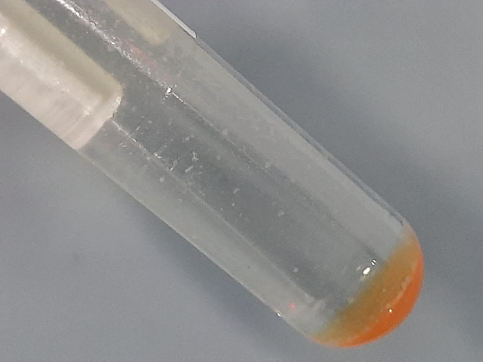
- The Appearance: As the MTBC grows, it forms granules, flakes, or “crumbs” that settle at the bottom of the tube. These aggregates are almost always off-white to buff in color.
- The Contrast: This pale, neutral color is a key diagnostic clue. If the sediment appears bright yellow, orange, or pink, it strongly suggests a scotochromogenic NTM (Non-Tuberculous Mycobacteria) or fungal contamination rather than M. tuberculosis.
Why does it look “Buff”
The color is a result of the high lipid content in the MTBC cell wall and the lack of carotenoid pigments. In a liquid MGIT:
- Cording: Because MTBC is hydrophobic and produces cord factor, it doesn’t stay suspended. It clumps together.
- Density: When these clumps gather at the bottom (especially after centrifugation or undisturbed incubation), the density of the mass makes the buff/cream color quite distinct against the orange-tinted sensor at the base.
Comparison: MTBC vs. Other Growth
| Growth Feature | M. tuberculosis (MTBC) | NTM / Contaminants |
| Color | Buff / Off-white | Can be bright yellow, orange, or white |
| Texture | Granular, “bread-crumb” like | Smooth, mucoid, or uniform turbidity |
| Liquid Clarity | Supernatant usually stays clear | Often becomes cloudy (turbid) |
Note: Always remember that while a buff-colored granular sediment is classic for MTBC, it is not definitive. A ZN stain to check for cording and an MPT64 antigen test are still the gold standards for confirmation once the tube flags positive.
Key Elements Included:
- MTBC: Specific identification of the pathogen complex.
- MGIT: Reference to the liquid growth indicator system.
- Buff Color: Highlighting the specific pigmentation discussed.
- Granular/Cording: Acknowledging the unique flaky texture of the growth.
Further Readings
- https://www.nptccd.health.gov.lk/wp-content/uploads/2022/03/Laboratory-Manual-for-TB-Control-5th-Edition.pdf
- https://dokumen.pub/diagnosis-of-mycobacterium-9819956234-9789819956234.html
- https://www.microbiologyresearch.org/content/journal/jmm/10.1099/jmm.0.000734?crawler=true&mimetype=application/pdf
- https://pubmed.ncbi.nlm.nih.gov/26618171/
- https://pmc.ncbi.nlm.nih.gov/articles/PMC5796708/
- https://www.zgflzz.cn/EN/Y2013/V35/I1/27
- https://www.annclinlabsci.org/content/33/2/179.full
- https://tbcindia.mohfw.gov.in/wp-content/uploads/2023/05/6995271860Training-manual-M-tuberculosis-C-DST.pdf
- https://www.jaypeedigital.com/eReader/chapter/9789386261359/ch1
- https://www.scielo.cl/scielo.php?script=sci_arttext&pid=S0716-97602017000100214