
Introduction
Table of Contents
Acinetobacter ursingii is a Gram-negative, non-fermentative, opportunistic bacterium belonging to the genus Acinetobacter. It is an uncommon species compared to Acinetobacter baumannii, but it has been increasingly recognized in healthcare-associated infections (HAIs). Reported cases include bacteremia, septicemia, pneumonia, urinary tract infections, and bloodstream infections, particularly in immunocompromised or hospitalized patients.
Morphology

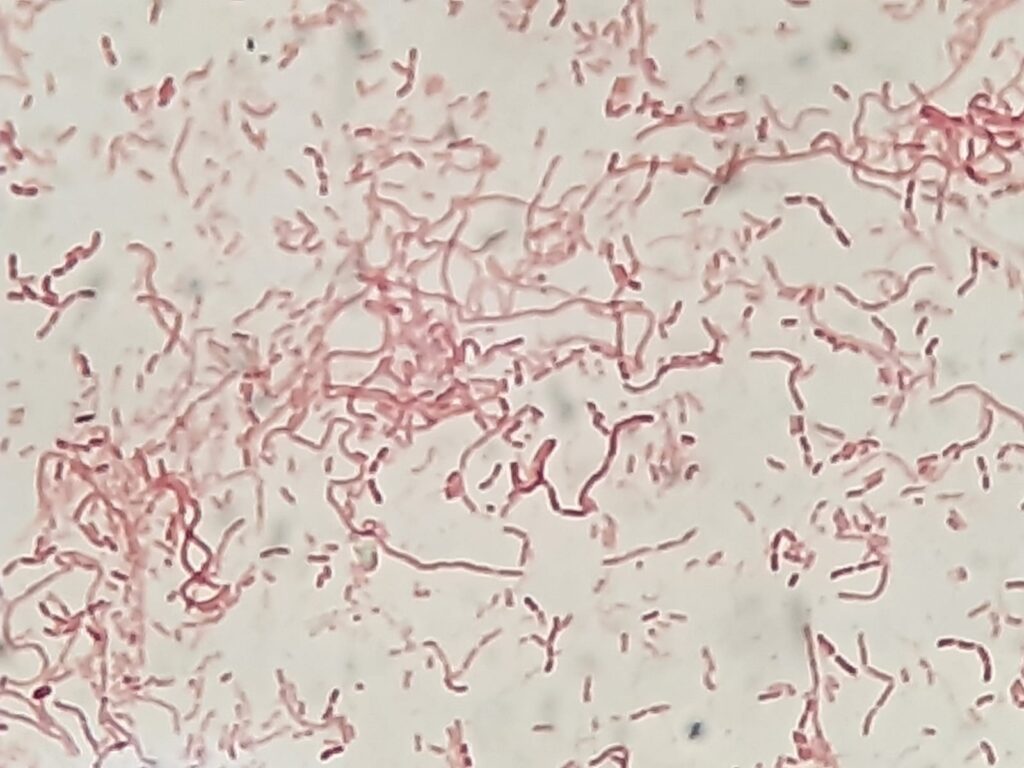
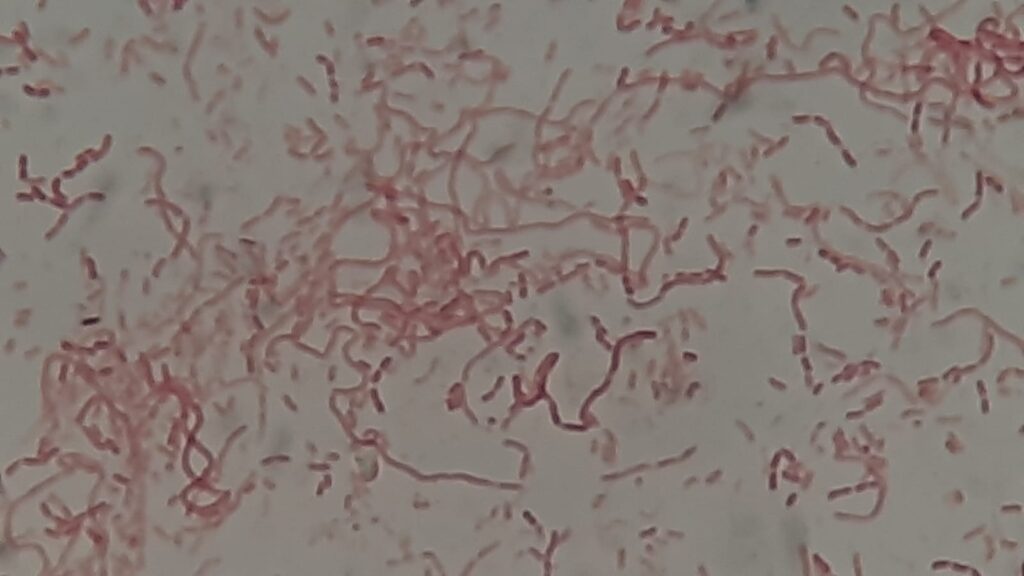
- Gram Stain: Gram-negative coccobacilli, often appearing as short rods.
- Growth on Media:
- Grows well on blood agar and MacConkey agar.
- Colonies: smooth, opaque, circular, and non-pigmented.
- Motility: Non-motile.
- Biochemical Features:
- Oxidase negative, catalase positive.
- Non-fermenter, utilizes limited carbon sources.


Pathogenicity
- Acts as an opportunistic pathogen, causing disease in patients with underlying conditions or indwelling devices.
- Clinical Manifestations:
- Bacteremia/septicemia (most common).
- Pneumonia (especially ventilator-associated).
- Urinary tract infections.
- Wound and catheter-associated infections.
- Produces biofilms, enhancing persistence in hospital environments and on medical devices.
- Emerging reports of multidrug resistance complicate management.
Laboratory Diagnosis

- Specimen Collection: Blood, sputum, urine, wound swabs, catheter tips.
- Culture: Growth on blood agar, MacConkey agar. Colonies may resemble other Acinetobacter spp.
- Identification:
- Conventional biochemical testing may misidentify the species.
- Accurate identification requires MALDI-TOF MS or molecular sequencing (16S rRNA, rpoB gene).
- Antimicrobial Susceptibility Testing (AST): Performed by CLSI/EUCAST broth microdilution or automated systems.

Treatment
- Most strains are susceptible to a wider range of antibiotics than A. baumannii, but resistance is increasing.
- Therapeutic options:
- Beta-lactams (piperacillin-tazobactam, cephalosporins, carbapenems in some cases).
- Fluoroquinolones, aminoglycosides, tetracyclines.
- Multidrug-resistant isolates: May require colistin or combination therapy.
- Therapy should always be guided by susceptibility testing.

Prevention

- Infection control measures in hospitals (hand hygiene, device care, surface disinfection).
- Judicious antibiotic use to prevent resistance selection.
- Active surveillance in ICUs and oncology wards.
- Strict sterilization of indwelling medical devices.
Keynotes

- Acinetobacter ursingii is a rare but emerging opportunistic pathogen in hospital settings.
- Causes primarily bloodstream and device-associated infections in immunocompromised patients.
- Often misidentified without molecular or MALDI-TOF confirmation.
- Treatment outcomes are generally better than for A. baumannii, but resistance is rising.
- Strong infection prevention practices are essential to reduce transmission.
Further Readings
- https://bmcinfectdis.biomedcentral.com/articles/10.1186/s12879-015-1145-z
- https://pmc.ncbi.nlm.nih.gov/articles/PMC8464211/
- https://pmc.ncbi.nlm.nih.gov/articles/PMC4907768/
- https://www.panafrican-med-journal.com/content/article/23/193/full/
- https://epi.utah.gov/wp-content/uploads/LTCF_Acinetobacter_FS.pdf
- https://www.cdc.gov/acinetobacter/about/index.html
- https://www.sciencedirect.com/topics/biochemistry-genetics-and-molecular-biology/acinetobacter
- https://journals.lww.com/md-cases/fulltext/2020/11000/community_acquired_acinetobacter_ursingii_occult.11.aspx
- https://iubmb.onlinelibrary.wiley.com/doi/10.1002/iub.534
- https://pubmed.ncbi.nlm.nih.gov/18197724/
- https://www.ezbiocloudpro.app/app/wiki/S;Acinetobacter%20ursingii
- https://pmc.ncbi.nlm.nih.gov/articles/PMC150291/
- https://www.researchgate.net/publication/10867688_Bacteremia_Caused_by_Acinetobacter_ursingii