
Introduction of Culture Media
Table of Contents
Culture media are required to grow the organisms and they are generally from clinical specimens to identify the causative agent.
Common Ingredients of Culture Media
Common Ingredients of Culture Media and their basic constituents are as follows-
Water: It is the source of hydrogen and oxygen.
Electrolyte: Sodium chloride or other electrolytes that maintain osmotic balance.
Peptone: It is a complex mixture of partially digested protein. Proteoses, amino acids, polypeptides, phosphates, minerals (K, Mg), and accessory growth factors like nicotinic acid and riboflavin are the constituents of peptone.
Meat extract: It is available commercially as “Lab-Lamco” which contains protein degradation products, inorganic salts, carbohydrates, and growth factors.
Blood or serum: It uses for enriching culture media. Usually, 5-10% defibrinated sheep blood is used for fastidious organisms like Streptococcus pneumoniae, Neisseria gonorrhoeae, and Neisseria meningitidis. Blood is also useful for chocolate agar for the recovery of another fastidious bacterium like H. influenzae. In a certain medium, the serum uses for the cultivation of organisms. e.g. Loeffler Serum slope and is used for the cultivation of Corynebacterium diphtheriae.
Agar: It is a long-chain polysaccharide prepared from sea wood (Algae –Geladium Species) that does not provide any nutrition to the microbes but acts as a solidifying agent only. Its common applicable concentration is 2-3%. Agar melts at 980C and solidifies at 420C. New Zealand agar has twice the capacity jellifying capacity as that of Japanese agar.
Types of Culture Media
Media are classified in flowing ways:
- Based on the physical state
- Liquid media
- Semisolid media ( Agar, 0.2-0.4% which enables motile bacteria to spread.)
- Solid media
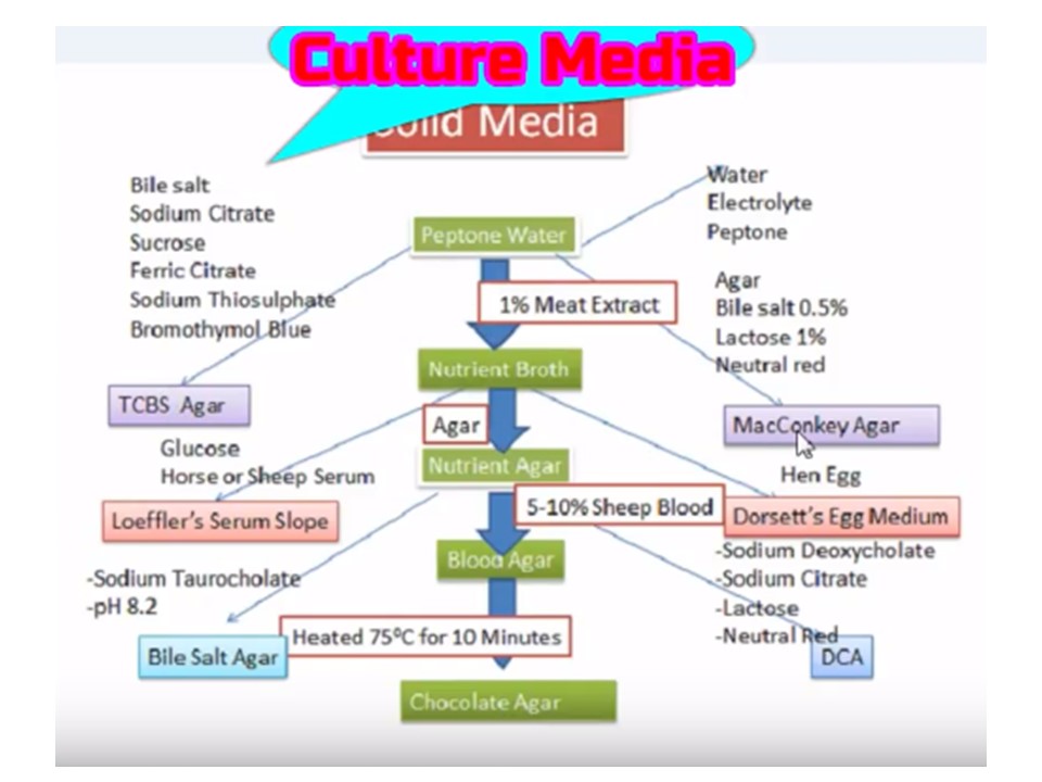
Liquid Culture Media Composition Flowchart

Solid Culture Media Composition Flowchart
- On the basis of the presence of molecular oxygen and reducing substances in the media
- Aerobic media
- Anaerobic media
- Based on nutritional factors
- Simple media
- Complex media
- Synthetic media
- Special media
Special media –
- Enriched media
- Enrichment media
- Selective media
- Differential media
- Indicator media
- Transport media
- Sugar media
Simple media: The nutrient broth is an example of a simple medium. It contains peptone water and meat extract of 1%. After the addition of agar to the nutrient broth, it becomes nutrient agar. This is the simplest and routinely employed medium in the laboratory for diagnostic purposes.
Complex media: All cultural media other than simple media are complex media.
Synthetic media: These are prepared from pure chemicals and the exact composition of the medium is known and those are used for special studies such as metabolic requirements. Dubo’s medium with tween 80 is an example of a synthetic medium.
Special Media
Enriched media: When a basal medium is added with some nutrients such as blood, serum, or egg that is called an enriched medium. For e.g. blood agar-Blood is added to nutrient agar that may be used for growing a number of bacteria but one specific example is Streptococcus which requires blood for its growth. Loeffler’s serum slope-Serum is added for enriching the medium. This medium is used for growing Corynebacterium diphtheriae.
Enrichment media: It is a fluid type of selective medium in which there is the incorporation of some substances that have either a stimulating effect on the bacteria to be grown or inhibits its competitors or both. This results in an absolute increase in the number of wanted bacteria related to other bacteria. Such a medium is called an enrichment medium. In tetrathionate broth, tetrathionate is added which inhibits coliforms while allowing typhoid-paratyphoid bacilli to grow. Similarly, in selenite F broth, Selenite has a similar action as that of tetrathionate in tetrathionate broth.
Selective media: Selective media contain substances that inhibit all but a few types of bacteria and facilitate the isolation of a particular species. These media are used to isolate a particular bacteria from a specimen where mixed bacterial flora is expected. Selective media are solid in contrast to enrichment media which are liquid. Examples of selective media are deoxycholate citrate agar(DCA)-The addition of deoxycholate acts as a selective agent for enteric bacilli (Salmonella, Shigella). Bile salt agar(BSA)-Bile salt is a selective agent that favors the growth of only Vibrio cholerae whereas inhibits the growth of other intestinal organisms.
Differential media: When a medium contains substances that help to distinguish differing characteristics of bacteria, it is called differential medium e.g. MacConkey’s medium, which contains peptone, lactose, agar, sodium taurocholate, and neutral red. The lactose fermenters (LF) form pink-colored colonies whereas non-lactose fermenters (NLF) produce colorless or pale colonies.
Indicator Media: These media contain an indicator that changes color when a bacterium grows in them. Salmonella enterica serotype Typhi grows as black colonies on Wilson and Blair’s medium containing sulfite. MacConkey ‘s medium is also an indicator medium. Due to the fermentation of lactose, there is acidic pH which forms the pink colonies in the presence of a neutral red indicator.
Transport media: These are used in the case of delicate organisms (e.g. gonococci) which may not survive the time taken for transit or may be overgrown by nonpathogenic bacteria(e.g. cholera organisms). They maintain only viability. Examples of transport media are Stuart’s transport medium-a non-nutrient soft agar gel containing a reducing agent to prevent oxidation, and charcoal to neutralize bacterial inhibitors. It may be used for organisms such as gonococci and Buffered glycerol saline transport medium for enteric bacilli.
Sugar media: Sugar media help in the identification of bacteria. The term sugar in microbiology denotes any fermentable substance. Glucose, lactose, sucrose, and mannitol are routinely employed for fermentation tests.
Anaerobic Media: These are used for the cultivation of anaerobic bacteria e.g.
- Cooked meat broth (CMB)
- Thioglycollate broth
Preparation and Uses of Common Culture Media
In this subtopic, common culture media like nutrient agar, blood agar, MacConkey agar, chocolate agar, and antimicrobial susceptibility testing (AST) medium i.e. Muller-Hinton agar (MHA) are discussed in detail.
Introduction of Nutrient Agar
Nutrient agar is a simple medium which uses to grow bacteria that is devoid of indicators, selective agents, differential ingredients, and enriching substances therefore uses for better expression of pigmentation, biochemical test, and even serotyping.

Composition of Nutrient Agar
| Constituents | Gm/Liter |
| Lab-Lemco’ powder | 1.0 |
| Yeast extract | 2.0 |
| Peptone | 5.0 |
| Sodium chloride | 5.0 |
| Agar | 15.0 |
| Distilled water | 1000 ml |
| pH | 7.4 ± 0.2 @ 25°C |
Principle of Nutrient Agar
Peptone is an enzymatic digest of animal protein and the principal source of organic nitrogen for growing bacteria. Lab-Lemco powder( beef extract) and yeast extract are water-soluble ingredients of nutrient agar that contribute to vitamins, carbohydrates, nitrogen, and salts. Sodium chloride maintains the osmotic equilibrium of the medium. The presence of sodium chloride in nutrient agar maintains a salt concentration that is similar to the cytoplasm of the microorganisms. Agar acts as the solidifying agent whereas water is an essential ingredient for the growth and reproduction of organisms. water also serves as a transport medium for the agar’s various substances.
Preparation of Nutrient agar
- Suspend 28.0 grams in 1 liter of purified/distilled or deionized water.
- Heat to boiling to dissolve the medium completely.
- Sterilize by autoclaving at 15 lbs pressure (121°C) for 15 minutes.
- After autoclaving, leave for cooling to 45-50°C.
- Pour nutrient agar into each plate and leave plates on the sterile surface until the agar has solidified.
- Store the plates in a refrigerator at 2-8°C.
Storage and Shelf life of Nutrient agar
- Store at 2-8ºC and away from direct light.
- Media should not be used if there are any signs of deterioration (shrinking, cracking, or discoloration), or contamination.
- The product is light and temperature sensitive; protects from light, excessive heat, moisture, and freezing.
Test Requirements
- Test specimens ( samples or growth of bacteria)
- Inoculating loop
- Bunsen burner
- Incubator
- Control strains (Escherichia coli ATCC 25922 and Staphylococcus aureus
ATCC 25923)
Test procedure (specimen/organism inoculation)
- Allow the plates to warm at 37°C or to room temperature, and the agar surface to dry before inoculating.
- Inoculate and streak the specimen as soon as possible after collection.
- If the specimen to be cultured is on a swab, roll the swab over a small area of the agar surface.
- Streak for isolation with a sterile loop.
- Incubate plates aerobically at 35-37ºC. for 18-24 hours.
- Examine colonial characteristics.
Result and Interpretation
Control strains: Escherichia coli ATCC 25922 and Staphylococcus aureus
ATCC 25923): good-luxuriant
Presence of non-fastidious bacteria in specimen: Presence of growth on nutrient agar
Uses of Nutrients Agar
- For the cultivation and maintenance of non-fastidious bacteria.
- Preparation of blood agar
- It is also used in antibiotic sensitivity testing
- Concentrated agar up to 3 more % prevents swarming of Proteus species as well as Clostridium tetani.
- Preparation of chocolate agar ( heating blood agar changes to chocolate agar).
- It uses for better expression of pigmentation.
- It is also used for serotyping of organisms.
- It also uses for the isolation of pure cultures from mixed growth.
- Nutrient agar is also beneficial for the enumeration of organisms in water, sewage, dairy products, feces, and other materials.
Keynotes
- Nutrient broth contains the same ingredients except for agar.
- Earlier it was used as blood and chocolate agar base medium but nowadays replaced by various manufacturers.
Limitations of Nutrient Agar
- Individual organisms differ in their growth requirement and may show variable growth patterns in the medium.
- Each lot of the medium has been tested for the organisms specified on the certificate of analysis. It is recommended to users validate the medium for any specific microorganism other than that mentioned in the certificate of analysis (COA) based on the user’s unique requirement.
- It is recommended that biochemical, immunological, molecular, or mass spectrometry testing be performed on colonies from pure culture for complete identification.
- It is a general-purpose medium and thus for recovering fastidious organisms like S. pneumoniae and H. influenzae nutrient agar should be modified.
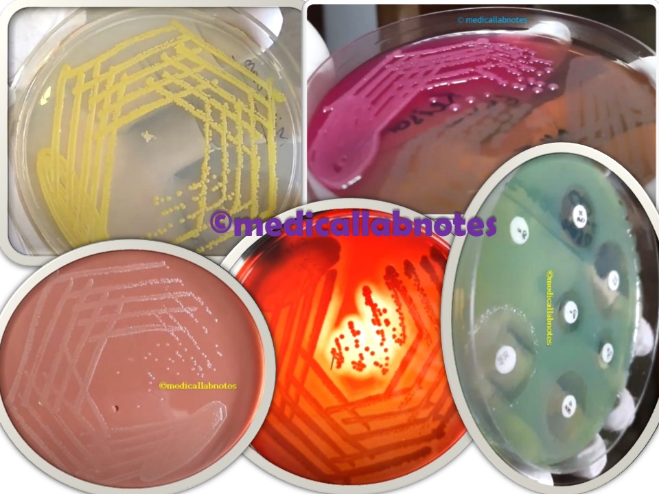
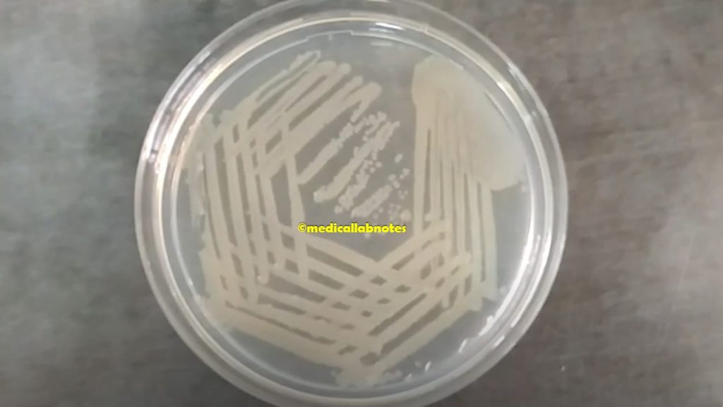
Nutrient Agar Footages
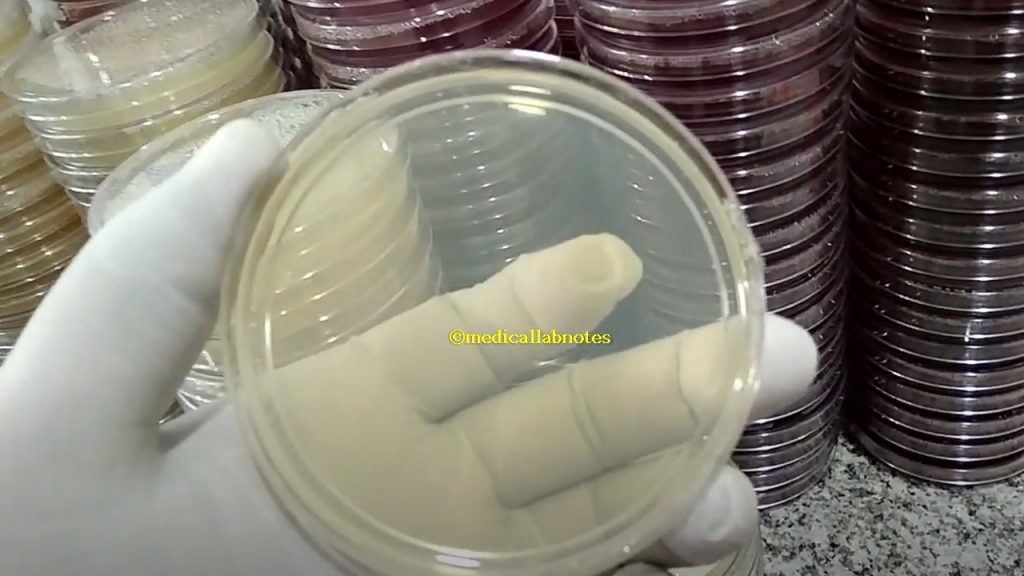
Staphylococcus aureus colony morphology on nutrient agar
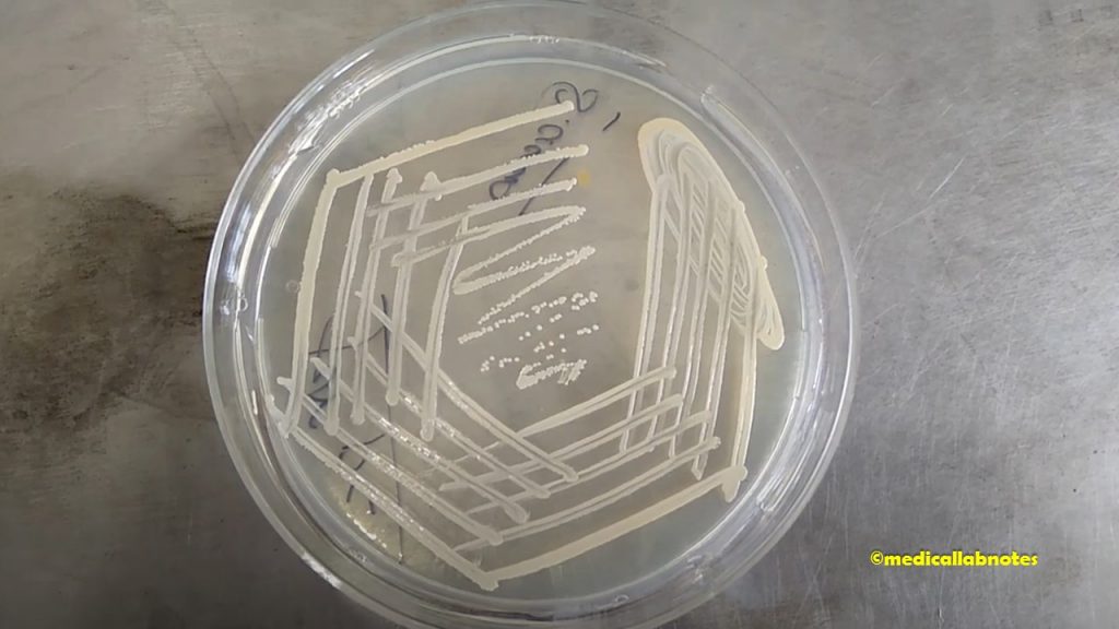
Micrococcus luteus colony morphology on nutrient agar
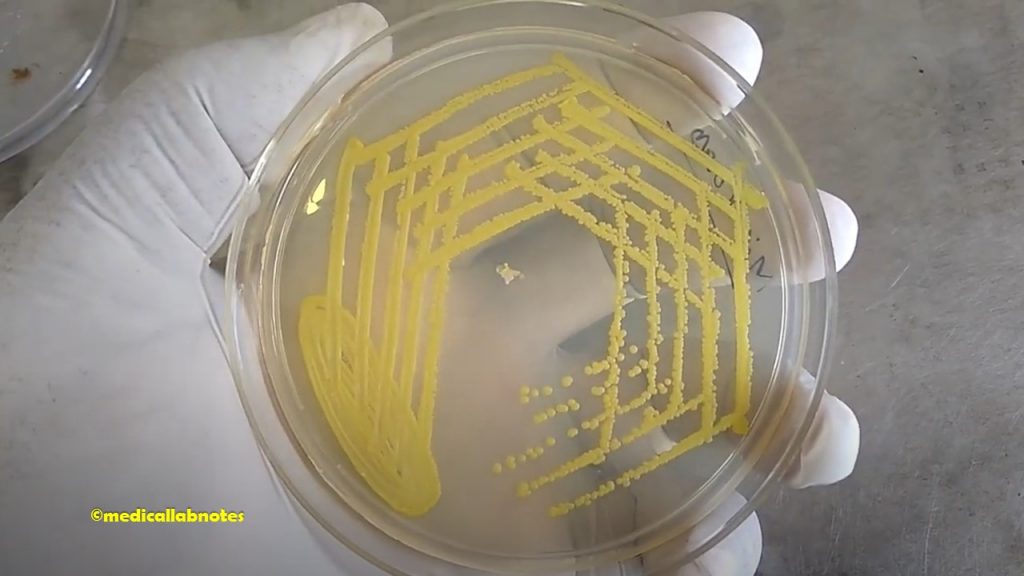
Vibrio cholerae colony characteristics on nutrient agar
Introduction of MacConkey Agar
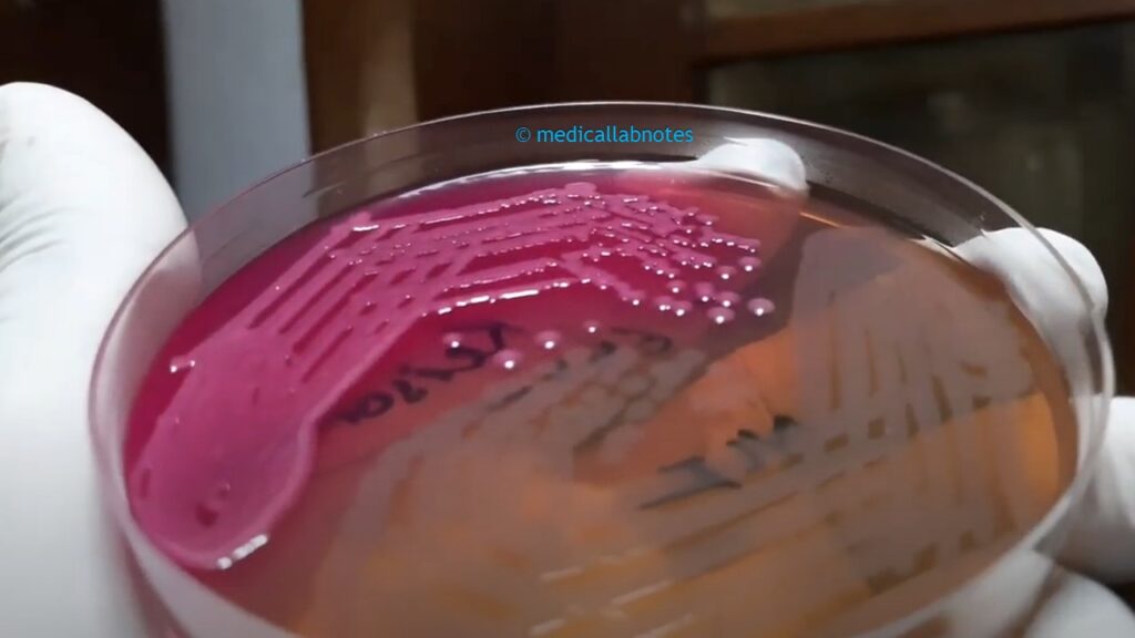
MacConkey agar (MAC) uses for the cultivation of gram-negative bacteria and this Enterobacteriaceae belonging bacteria grow well on this medium. Coliforms also enjoy this medium and MacConkey Agar is a modification of Neutral Red Bile Salt Agar. MacConkey developed it. It was one of the earliest culture media for the cultivation and identification of enteric organisms from clinical specimens as well as food and water. It is a selective, differential, and indicator medium because of the following properties-
- Selective due to bile salts that inhibit gram-positive bacteria and select gram-negative bacilli.
- The indicator medium is due to having neutral red incorporated in it.
- Differential medium is due to separate whether lactose fermenter or non-lactose fermenter bacteria.
- The above picture is showing the lactose and non-lactose fermenter colony of bacteria.
Principle of MacConkey Agar
The recommendation of MacConkey Agar is as a selective and differential medium for the isolation of gram-negative bacilli including coliform organisms and enteric pathogens, on the basis of lactose fermentation. Peptones (meat and casein) and pancreatic digest gelatin and provide the essential nutrients, vitamins, and nitrogenous factors required for the growth of microorganisms. Lactose monohydrate is a fermentable source of carbohydrates. The attribution of the selective action of this medium is due to crystal violet and bile salts, which are inhibitory to most species of gram-positive bacteria. Sodium chloride maintains the osmotic balance in the medium here as neutral red is a pH indicator that turns red at a pH below 6.8 and is colorless at any pH greater than 6.8. Agar is the solidifying agent.
Composition of MacConkey Agar
(Himedia)
| Ingredients | Gms / Litre |
| Peptones (meat and casein) | 3.0 |
| Pancreatic digest of gelatin | 17.0 |
| Lactose monohydrate | 10.0 |
| Bile salts | 1.5 |
| Sodium chloride | 5.0 |
| Crystal violet | 0.001 |
| Neutral red | 0.03 |
| Agar | 13.5 |
| Distilled water | 1000 ml |
| pH after sterilization( at 25°C) | 7.1±0.2 |
Preparation of MacConkey agar
- Suspend 49.53 grams of the dehydrated medium in 1000 ml of purified/distilled water.
- Heat to boiling to dissolve the medium completely.
- Sterilize by autoclaving at 15 lbs pressure (121°C) for 15 minutes i.e. validated cycle.
- Cool to 45-50°C.
- Mix well before pouring into sterile Petri plates.
- Leave for drying.
Storage and Shelf life of MacConkey Agar
- Store at 2-8ºC and away from direct light.
- Media should not be used if there are any signs of deterioration (shrinking, cracking, or discoloration), or contamination.
- The product is light and temperature-sensitive; protects from light, excessive heat, moisture, and freezing.
Test procedure (specimen/organism inoculation)
- Allow the plates to warm at 37°C or to room temperature, and the agar surface to dry before inoculating.
- Inoculate and streak the specimen as soon as possible after collection.
- If the specimen to be cultured is on a swab, roll the swab over a small area of the agar surface.
- Streak for isolation with a sterile loop.
- Incubate plates aerobically at 35-37ºC. for 18-24 hours.
- Examine colonial characteristics.
Colony Characteristics of various organisms in MacConkey Agar
Lactose-positive (pink colonies): Lactose fermenting species will grow pink colonies. Lactose fermentation will produce acidic by-products that lower the pH and this turns the pH indicator pink. e.g. Escherichia coli, Enterobacter, Klebsiella while lactose-negative (white colonies): Gram-negative bacterial species will still form colonies, but colonies will have a white appearance as there will be no change in pH in the absence of lactose fermentation. e.g. Salmonella, Proteus, Yersinia, Pseudomonas
No colonies: Gram-positive bacteria will not form any colonies on the MacConkey medium. e.g. Staphylococcus, Enterococcus, Micrococcus
Slow or weak Lactose positive: Weak lactose fermenters will form colonies slower than the rest. e.g. Serratia, Citrobacter
Mucoid (sticky, wet colonies): Encapsulated bacteria produce capsules using lactose. This gives sticky, wet-appearing colonies and mucoid colony-forming species are Klebsiella, and Enterobacter.
Keynotes:
It is of various types on the purpose of uses like-
- MacConkey agar without bile salt- It uses both gram-negative and gram-positive bacteria
- McConkey agar with bile salt- Selective for gram-negative bacteria but Enterococcus species may grow.
- MacConkey agar with bole sat and crystal violet: Strict selective medium for gram-negative bacteria that also inhibits Entercoccus species due to having crystal violet in its composition.
- The amount of medium for preparation also varies slightly from manufacturer to manufacturer. e.g. Himedia 49.53 gm for 1 liter, whereas Oxoid 51.5 gm and Hardy Diagnostics 52.49 gm.
- MacConkey Agar with Sorbitol is to be used as a selective and differential medium for the detection of enterohemorrhagic Escherichia coli O157:H7.
Uses of MacConkey agar (MAC)
- MacConkey Agar is recommended for use as a selective, differential, and indicator medium for the isolation of gram-negative bacilli including coliform organisms and enteric pathogens.
- It is used in the differentiation of lactose fermenting from lactose non-fermenting gram-negative bacteria.
- It is used for the isolation of coliforms and intestinal pathogens from clinical specimens as well as food and water samples.
Limitations of MacConkey Agar
- Colony characteristics only provide presumptive identification and thus biochemical, immunological, molecular, or mass spectrometry testing be performed on colonies from pure culture for final identification.
- The concentration of bile salts in MacConkey Agar is relatively low in comparison with other enteric plating media. The parallel use of more selective media for gram-negative enterics, such as Hektoen enteric agar (HEK, HE, or HEA)or Xylose lysine deoxycholate (XLD) agar is recommended in order to increase the chances of pathogen isolation.
- Some strains of the organism may be encountered that grow poorly or fail to grow on this medium
- Some strains of Proteus may swarm on this medium.
- Serial inoculation may be required to assure adequate isolation of mixed flora samples.
- Incubation of MacConkey Agar plates under increased CO2 has been reported to reduce the growth and recovery of a number of strains of Gram-negative bacilli.
Introduction of Blood Agar
Blood agar is an enriched medium for the cultivation of bacteria. Fastidious organisms like streptococci, do not grow well on ordinary growth media. It is a type of growth medium i.e. trypticase soy agar enriched with 5% sheep blood or blood agar base with 5-10 % sterile sheep blood. Medium encourages the growth of bacteria and their hemolysis, such as streptococci, that otherwise wouldn’t grow.

Composition of Sheep Blood Agar Base
| Ingredients | Gms / Litre |
| Casein enzymic hydrolysate | 14.0 |
| Peptic digest of animal tissue | 4.5 |
| Yeast extract | 4.5 |
| Sodium chloride | 5.0 |
| Agar | 12.5 |
| Final pH (at 25°C) | 7.3±0.2 |
Procedure for the Preparation of Blood Agar
- Suspend 40.5 grams in 1000 ml of distilled water or deionized water.
- Heat to boiling to dissolve the medium completely.
- Sterilize by autoclaving at 15 lbs pressure (121°C) for 15 minutes.
- Cool to 45-50°C and aseptically add 7% sterile sheep blood.
- Mix well and pour into sterile Petri plates. Avoid the formation of air bubbles. You must have warmed the blood to room temperature at the time of dispensing it to a molten agar base.
- Dispense 15 ml amounts to sterile Petri plates aseptically
- Label the medium with the date of preparation and give it a batch number (if necessary).
Storage and Shelf Life
Store below 30°C in a tightly closed container and the prepared medium at 2-8°C, preferably in sealed plastic bags to prevent loss of moisture. The shelf life of this prepared blood agar is up to four weeks. Use it before the expiry date on the label.
Principle and Interpretation of Blood Agar
Hemolysins are exotoxins that bacteria produce that lyse red blood cells. The hemolytic reaction can be visualized on blood agar plates observed through the bright transmitted light. On blood agar plates colonies of hemolytic bacteria may be surrounded by a clear, colorless zone where the red blood cells have been lysed and the hemoglobin destroyed to a colorless compound and which is beta hemolysis. Other types of bacteria can reduce hemoglobin to methemoglobin which produces a greenish zone around the colonies and is called alpha hemolysis. Gamma hemolysis is lacking hemolysis where no change in the medium is observed.
Sheep blood agar base with added sheep blood was developed to allow maximum recovery of organisms without interfering with their hemolytic reactions. The sheep blood agar base was formulated to be compatible with sheep blood and give improved hemolytic reactions of organisms. Casein enzymic hydrolysate and yeast extract provide nitrogen, carbon, amino acids, and vitamins. Peptic digest of animal tissue (PDAT) is the nitrogen source. Sodium chloride (NaCl) maintains the osmotic balance. Sheep blood agar base showed considerable improvement and the expected beta-hemolytic reactions with S. pyogenes in comparison to other blood agar bases supplemented with blood
Quality control
- Inoculate Streptococcus pneumoniae and Streptococcus pyogenes into a prepared blood agar.
- Observation of Cultural characteristics after incubation at 35-37°C for 18-24 hours with added 7% v/v sterile sheep blood.
| Organisms | Growth | Hemolysis |
| Streptococcus pneumoniae ATCC 6303 | luxuriant | alpha |
| Streptococcus pyogenes ATCC 19615 | luxuriant | beta |
- This is an indicator of good quality control of prepared blood agar.
Uses of Blood Agar
Blood agar uses for the following purposes-
- Determine the type of hemolysis, whether alpha, beta, or gamma.
- For the culture of streptococci as well as antimicrobial susceptibility testing (AST).
- Use of optochin disc for presumptive identification of Streptococcus pneumoniae.
- Similarly use of bacitracin disc (0.04U) for presumptive identification of Streptococcus pyogenes.
- To perform the CAMP test for Streptococcus agalactiae.
- To perform satellitism test for Haemophilus.
- It is also used for the isolation and cultivation of other than streptococci like Neisseria and other fastidious microorganisms.
- It is also useful for the preparation of Salmonella Typhi antigens.
Keynotes
- Blood is a good constituent of enriched medium for fastidious organisms even though contains inhibitors for certain bacteria such as Neisseria and Haemophilus genera and the blood agar must be heated to inactivate these inhibitors and to release essential growth factors (e.g., V factor). The heating of blood agar converts it into chocolate agar (75°C for 15 minutes turns a chocolate color) and supports the growth of these bacteria.
- Hemolysis on blood agar: Mainly three types of hemolysis are produced in Sheep blood agar by Streptococci namely; Alpha hemolysis, Beta hemolysis, and gamma hemolysis but sometimes alpha prime or wide zone alpha hemolysis may be encountered. Hemolysis is best observed by examining colonies grown under anaerobic conditions or inspecting sub-surface colonies. How does one know if the colonies they are observing on a plate have caused alpha hemolysis or beta hemolysis or gamma ?-Follow up principle and interpretation.
- To check the type of blood agar hemolysis, the blood agar plate must be held up to a bright transmitted light source and observed with the light coming from behind.
- Alpha hemolysis: Streptococcus pneumoniae
- Beta Hemolysis: Group A beta-hemolytic streptococci-Streptococcus pyogenes and Group B, beta-hemolytic streptococci–Streptococcus agalactiace. For the group, streptococci maximal activity of both the hemolysins; Oxygen labile SLO, and oxygen stable SLS hemolysins is observed only in anaerobic conditions.
- Gamma or non-hemolysis: Enterococcus species
- Alpha prime or wide zone alpha hemolysis: A small zone of intact erythrocytes immediately adjacent to bacterial colony, with a zone of complete red-cell hemolysis surrounding the zone of intact erythrocytes. This type of hemolysis may be confused with Beta hemolysis.
- The double zone of hemolysis on it is also seen by some organisms like Clostridium perfringens and Aeromonas hydrophilia called target hemolysis.
- If you are planning to prepare a batch of blood agar plates, prepare a few blood agar plates first to ensure that the blood is sterile.
Blood Agar Footages
Staphylococcus aureus beta-hemolytic colony on blood agar
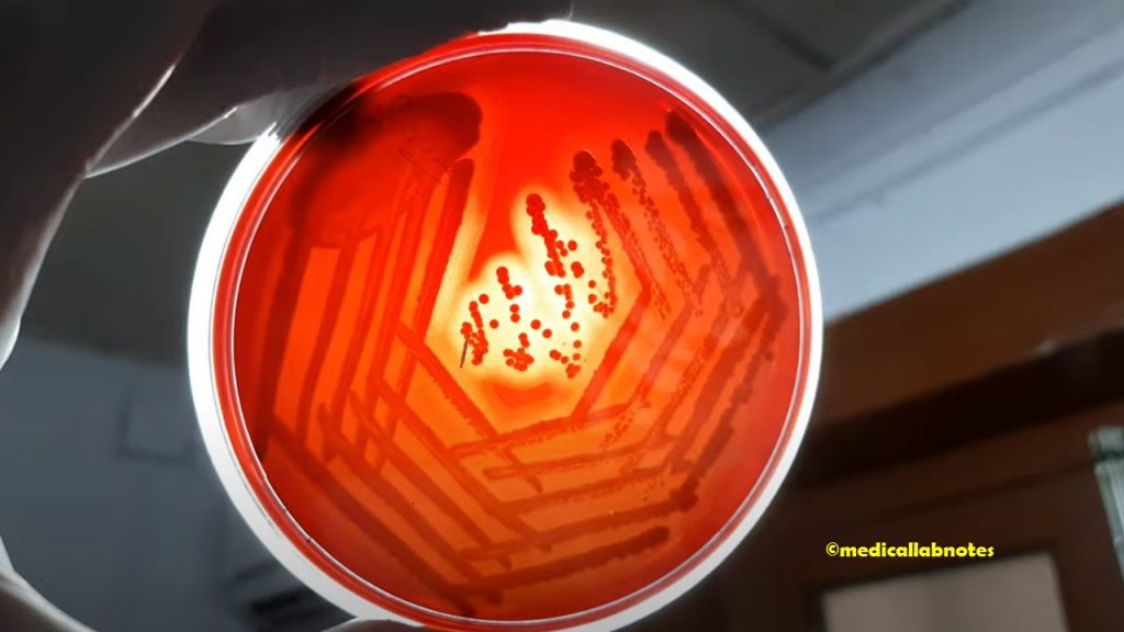
Streptococcus pneumoniae Draughtsman colony on 5% sheep blood agar

Streptococcus agalactiae colony morphology on 5% sheep blood agar

Streptococcus pyogenes beta-hemolytic colony morphology on 5% sheep blood agar
Enterococcus colony morphology on blood agar showing gamma hemolysis

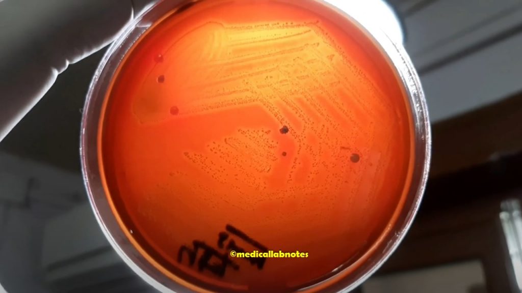
Chocolate Agar’s short form is (CHOC) and it is a non-selective, enriched growth medium that is the lysed blood agar. The name of the medium for its color is when the red blood cells (RBCs) lysis gives the medium a chocolate-brown color without having chocolate products. It uses for the isolation of fastidious bacteria, such as Haemophilus influenzae, when incubated at 35-37°C in a 5% CO2 incubator.

Composition of Blood Agar Base
| Ingredients | Gms / Litre |
| Casein enzymic hydrolysate | 14.0 |
| Peptic digest of animal tissue | 4.5 |
| Yeast extract | 4.5 |
| Sodium chloride | 5.0 |
| Agar | 12.5 |
| Final pH (at 25°C) | 7.3±0.2 |
Principle of Chocolate Agar
The composition of chocolate agar is the same as that of blood agar. The only difference of chocolate agar during preparing the red blood cells is lysed changing the medium color to chocolate brown. The lysis of RBC during the heating process releases intracellular coenzyme nicotinamide adenine dinucleotide (Factor V or NAD) into the agar for utilization by fastidious bacteria (the heating process also inactivates growth inhibitors). Hemin (factor X) is available from non-hemolyzed as well as hemolyzed blood cells. The most common species that require this enriched medium for growth include Neisseria meningitidis and Haemophilus spp. H. influenzae is not able to grow on sheep blood agar.
Requirements for Chocolate Agar Preparation
- Prepared blood agar
- Incubator for the simplest method
- But other methods are
- Blood agar base
- Sheep blood
- Distilled water
- Measuring cylinder
- Autoclave
- Weighing balance
- Water bath
- Hot air oven (optional)
Preparation of Chocolate Agar
It can be prepared by the following methods.
Simplest method
- Take already prepared blood agar plates (5% sheep blood agar) and put those plates into a hot air oven for 2 hours at 55°C.
- Take out those plates and you will get chocolate agar.
- Place the plates in sterile plastic bags and store them at 4°C until use.
- As a sterility test, incubate an uninoculated plate for 48 hours at 35-37°C with 5% CO2.
Another Method
- Suspend 40.5 grams in 1000 ml of distilled water or deionized water.
- Heat to boiling to dissolve the medium completely.
- Sterilize by autoclaving at 15 lbs. pressure (121°C) for 15 minutes.
- Heat-lyse a volume of sheep blood that is 5% of the total volume of media being prepared very slowly to 56°C in a water bath.
- Dispense 20 ml into 15×100 mm Petri dishes. Allow the media to solidify and condensation to dry.
- Place the plates in sterile plastic bags and store them at 4ºC until use.
- As a sterility test, incubate an uninoculated plate for 48 hours at 35-37°C with 5% CO2 (or in a candle jar).
Quality Control
For quality control, inoculate N. meningitidis, S. pneumoniae, and H. influenzae QC strains into prepared chocolate agar (CHOC) for 18-24 hours at 35-37°C with 5% CO2 (or in a candle jar but it can only provide up to 3% CO2).
| Organisms | growth |
| Neisseria meningitidis ATCC 13090 | luxuriant |
| Streptococcus pneumoniae ATCC 6303 | luxuriant |
| Haemophilus influenzae ATCC 19418 | luxuriant |
Storage and Shelf life of Chocolate agar
- Store at 2-8ºC and away from direct light.
- Media should not be used if there are any signs of deterioration (shrinking, cracking, or discoloration), or contamination.
- The product is light and temperature sensitive; protects from light, excessive heat, moisture, and freezing.
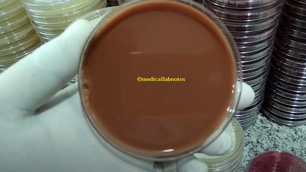
Freshly prepared chocolate agar without inoculation
Test Requirements
- Test specimens (Blood, vaginal samples, sputum, or growth of bacteria)
- Inoculating loop
- Bunsen burner
- Incubator
- Chocolate agar
- Control strains
Test procedure (specimen/organism inoculation)
- Allow the plates to warm at 37°C or to room temperature, and the agar surface to dry before inoculating.
- Inoculate and streak the specimen as soon as possible after collection.
- If the specimen to be cultured is on a swab, roll the swab over a small area of the agar surface.
- Streak for isolation with a sterile loop.
- Incubate plates aerobically at 35-37ºC for 18-24 hours with 5% CO2.
- Examine colonial characteristics.
Result -Interpretation
- Control strains i.e. Neisseria meningitidis ATCC 13090 and Haemophilus influenzae ATCC 19418: Good-luxuriant
- Test Organisms: Colony morphology depends on the nature of the organisms.
Colony Characteristics in Chocolate Agar
- Haemophilus influenzae: Non-hemolytic, opaque cream to gray colonies.
- Neisseria meningitidis: Growth on chocolate agar is grayish, non-hemolytic, round, convex, smooth, moist, glistening colonies with a clearly defined edge.
- Neisseria gonorrhoeae: Colonies on CHOC are pinkish-brown and translucent, exhibit smooth consistency and defined margins, and are typically 0.5-1 mm in diameter.
Uses of Chocolate Agar
- It is a very useful medium to isolate fastidious organisms in Microbiology laboratories from various clinical specimens like sputum (H. influenzae), urethral discharge (N. gonorrhoeae), and CSF/blood (N. meningitidis).
- And thus, Chocolate agar uses to isolate and cultivate fastidious microorganisms such as Haemophilus species and Neisseria species.
- It is also useful in isolating N. gonorrheae from both acute and chronic cases of gonococcal infections.
- It is also useful in isolating N. meningitidis from bacterial meningitis.
- Chocolate agar with bacitracin acts as a selective medium for screening H. influenzae from specimens e.g. sputum containing a mixed flora of microorganisms.
- Its modified forms use given below.
Modification of Chocolate Agar (CHOC)
- Thayer-Martin agar/medium uses for the selective isolation of N. gonorrhoeae and N. meningitidis. This Media is a chocolate agar supplemented with vancomycin, colistin, and nystatin (VCN) to inhibit the normal flora, including non-pathogenic Neisseria from the clinical specimens
- Chocolate Agar with bacitracin: CHOC with bacitracin is a selective medium used to improve the primary isolation of Haemophilus influenzae from specimens containing a mixed flora of microorganisms.
- Chocolate agar with GC base and growth supplement: It is a medium that supports the special growth requirements (hemin and NAD) needed for the isolation of fastidious organisms, such as H. influenzae, when incubated at 35-37°C in a 5% CO2 atmosphere.
- Chocolate agar with TSA and growth supplements: It is a medium that supports the special growth requirements (hemin and NAD) needed for the isolation of fastidious organisms, such as H. influenzae, when incubated at 35-37°C in a 5%CO2atmosphere.
Keynotes on Chocolate agar
- Chocolate agar is a recommended medium for the isolation of Neisseria gonorrhoeae from chronic and acute cases of gonococcal infections as well as H. influenzae from the sputum of upper respiratory tract infection.
- It is the medium of choice for the cultivation of fastidious bacteria.
- The organisms which grow on MacConkey medium and blood agar also grow on chocolate agar but not vice versa.
- Thayer-Martin agar, chocolate agar with bacitracin, chocolate agar with GC base and growth supplement, and chocolate agar with TSA and growth supplements are modified forms of chocolate agar.
Chocolate Agar Footages
Campylobacter colony morphology on chocolate agar
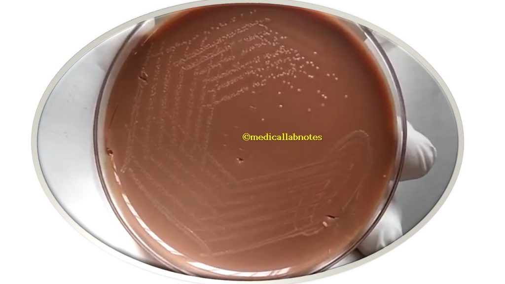
Haemophilus influenzae growth around bacitracin disk in chocolate agar of sputum culture

E. coli colony morphology on chocolate agar

Introduction of Muller-Hinton Agar
The name Mueller- Hinton agar(MHA) is from the surname of co-developers microbiologist John Howard Mueller and veterinary scientist Jane Hilton at Harvard University as a culture for gonococcus and meningococcus, who published the method in 1941. It is a non-selective and non-differential medium. In 1966, Bauer et al. adopted MHA for antimicrobial susceptibility testing. Later, Muller- Hinton agar (MHA) was adopted as the common medium to use for routine antibiotics susceptibility testing for non-fastidious bacteria (both aerobes and facultative anaerobes) that also used to isolate and maintain Neisseria and Moraxella species.
The use of media other than Mueller-Hinton agar may result in erroneous results. Now is recommended by various institutions like the American Society of Microbiology (ASM), Clinical, Laboratory Standards Institute (CLSI), and FDA, World Health Organization (WHO) for antibiotic susceptibility testing of bacteria.
Composition of MHA
| Ingredients | Gms / Litre |
| Beef infusion solids | 2.0 |
| Starch | 1.5 |
| Casein hydrolysate | 17.5 |
| Agar | 17.0 |
| Distilled water (D/W) | 1000 ml |
| Final pH at 25°C | 7.3 +/- 0.2 |
Principle of MHA
Ingredients of MHA are beef infusion solids, starch, casein hydrolysate, and agar. Beef infusion solids and casein hydrolysate provide nitrogen, vitamins, carbon, amino acids, sulfur, and other essential nutrients. Starch act as a “protective colloid” that absorbs any toxic metabolites produced in the medium. Starch hydrolysis yields dextrose, which serves as a source of energy whereas agar is the solidifying agent.
Properties of MHA
Due to the following properties, Muller- Hinton agar uses for routine antibiotic sensitivity testing (AST)-
- Defined medium concentration.
- Identical from batch to batch.
- No enrichment or selective in nature.
- It supports the growth of nearly all types of pathogens.
- Constituents are not antagonistic to any drugs.
- Well-adjusted pH(7.3) for all types of antibiotic susceptibility tests.
- The Agar-broth version has the same formula.
- Isotonic
- Appropriate for adding blood and serum.
- Adjusted mineral salts.
- It contains starch and it is known to absorb toxins released from bacteria so that they cannot interfere with antibiotics.
Note: Mueller- Hinton agar (MHA) is available in the market by commercial suppliers both ready-made form and dehydrated medium. Be sure to prepare the media according to the manufacturer’s directions.
Requirements for Preparation of Muller- Hinton Agar
- Dehydrated Mueller-Hinton agar powder
- Electronic balance
- Distilled water
- Autoclave
- Measuring cylinder
- Conical flask
- Hot plate
- Petri plates or dish
- Sheep blood ( optional for Mueller Hinton blood agar and Mueller Hinton chocolate agar)
- Control strains for testing the quality of prepared media
Preparation of MHA
- Suspend 38 g of medium in 1 liter of distilled water and mix thoroughly.
- Heat with frequent agitation and boil for 1 minute to completely dissolve the components.
- Autoclave at 121°C for 15 minutes.
- Cool to 45°C
- Pour cooled Mueller Hinton Agar into sterile Petri dishes on a level, horizontal surface to give uniform depth.
- Check the prepared Mueller Hinton agar to ensure the final pH is 7.3 ±1 at 25°C.
- Prepared media can be stored at 4 – 8°C. Mueller-Hinton agar is stable for approximately 70 days from the date of preparation but should be protected from direct light.
Storage and Shelf life of MHA
- Store at 2-8ºC and away from direct light.
- Media should not be used if there are any signs of deterioration (shrinking, cracking, or discoloration), or contamination.
- The product is light and temperature sensitive; protects from light, excessive heat, moisture, and freezing.
Test Requirements
- Pure cultures of the organism isolated from the clinical specimen
- Muller Hinton Agar
- Antibiotic Disks
- Turbidity Standard
- Swabs
Test procedure (specimen/organism inoculation)
- Mostly Muller Hinton agar( MHA) is used in this antibiotic susceptibility test.
- Emulsify 2-3 colonies in sterile saline matching the turbidity that standard (0.5 McFarland).
- Place a sterile cotton swab in the bacterial suspension and remove the excess fluid by pressing and rotating the cotton against the inside of the tube above the fluid level.
- The swab is streaked in three directions over the surface of the MHA to obtain uniform growth.
- Allow the plates to dry for 10-15 minutes.
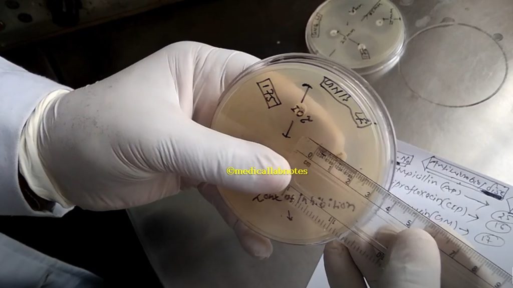
- Using sterile forceps or a suitable disk dispenser, place paper disks impregnated with a fixed concentration of an antibiotic, on the surface agar plates having a distance disc to disc 25 mm while the plate border to disk 15 mm.
- Incubate the plates at 37°C for 24 hours.
- Following overnight incubation, measure the diameter of the zone of inhibition in millimeters (mm) around each disc.
Result -Interpretation
- Using a standard table of antibiotic susceptibilities, determine if the strain is resistant, intermediate, or susceptible to the antibiotics tested.
Modifications of MHA
- Mueller Hinton agar medium supplemented with 5% sheep blood and nicotinamide adenine dinucleotide (NAD) is recommended for determining the antimicrobial susceptibility of Streptococcus species, Neisseria, and Campylobacter.
- Haemophilus test medium (HTM) is the preferred medium for the AST of H. influenzae using modified Kirby Bauer disc diffusion. HTM medium consists of the following ingredients: thymidine-free MHA supplemented with 15 μg/ml NAD, 15 μg/ml bovine hemin, and 5 mg/ml yeast extract.
- Mueller Hinton chocolate agar: For Haemophilus influenzae
- Mueller Hinton Agar No. 2: Thymine and thymidine inhibit sulfonamide and trimethoprim activity and calcium and magnesium interfere with the activity of aminoglycoside antibiotics. To overcome this problem MHA No. 2 is manufactured to contain low levels of thymine, and thymidine, and controlled levels of calcium and magnesium.
Quality Control
- Prepared Appearance: Light yellow to amber-colored clear to very slightly opalescent gel forms in Petri plates.
- Cultural Response: Observe cultural characteristics after incubation at 35°C-37°C for 18-24 hours.
| Organism | Growth |
| Escherichia coli (ATCC 25922) | Good |
| H. influenzae (ATCC 49247) | Good (on Mueller Hinton chocolate agar) |
| Neisseria gonorrhoeae (ATCC 49226) | Good |
| Pseudomonas aeruginosa (ATCC 27853) | Good |
| Staphylococcus aureus (ATCC 25923) | Good |
| Enterococcus faecalis (ATCC 29212) | Good |
| S. pneumoniae (ATCC 6305) | Good (on Mueller Hinton blood agar) |
Table: Control strains growth status
Other Media Uses for AST
- Bacteria: Aerobic and facultative anaerobes -Sensitest agar ( most common use outside of USA), Diagnostic sensitivity agar.
- Wilkins-Chalgren agar is recommended for testing anaerobic bacteria.
- For antiviral susceptibility testing, RPMI 1640 medium is used.
- Fungal susceptibility testing media are RPMI 1640 and Muller Hinton Agar + 2% Glucose and 0.5 mg/ml Methylene Blue Dye.
- Mycobacterium tuberculosis sensitivity testing media are Middle brook 7H- 9 ( broth + glycerol), 10 and 11 agar form.
Applications of MHA
- MHA is the common medium to use for routine antibiotics susceptibility testing for non-fastidious bacteria (both aerobes and facultative anaerobes).
- It is the standard medium for AST by the Bauer-Kirby method and its performance is specified by the CLSI.
- It is also an applicable medium for antimicrobial susceptibility testing by Stoke’s method.
- It can be used to cultivate Neisseria and Moraxella species.
- It is also a useful medium for the Epsilometer test (E-test) for measuring the MIC of the bacterial isolate.
- Antimicrobial susceptibility testing of fastidious bacteria can be tested using modified MHA like Mueller Hinton blood agar (for S. pneumoniae) and Mueller Hinton chocolate agar(for H. influenzae).
- Mueller Hinton agar is also specified in FDA Bacteriological Analytical Manual for food testing, and procedures commonly performed on aerobic and facultative anaerobic bacteria.
- Muller Hinton Agar + 2% Glucose and 0.5 mg/ml Methylene Blue Dye (GMB) Medium: It uses for antifungal susceptibility testing (AFST) of yeasts ( for Candida species).
- HiCrome™ Mueller Hinton Agar (for Antifungal Testing): Chromogenic differentiation of yeast cells along with antifungal susceptibility.
Limitations of MHA
- It is recommended medium for susceptibility testing of pure cultures only.
- Inoculum density may affect the size of the microbial growth zone of inhibition. Heavy inoculum may result in smaller zones or too less inoculum may result in larger zones.
- Fastidious organisms (S. pneumoniae, H. influenzae ) may not grow on this medium and may require the supplementation of blood.
- Fastidious anaerobes may not grow on this medium.
- As antimicrobial susceptibility testing is carried out with the antibiotic disc, proper storage of the disc is desired which may affect its potency of the disc.
Some keynotes on MHA
- The plates must be poured to a depth of 4 mm (approximately 25 ml of liquid agar for 100-mm plates and 60 ml of liquid agar for 150-mm plates, but in any case to a measured depth of 4 mm). Plates that are too shallow will produce false susceptible results as the antimicrobial compound will diffuse further than it should, creating larger zones of inhibition. Conversely, plates poured to a depth >4 mm will result in false resistant results.
- If the pH is <7.2 certain drugs will appear to lose potency (aminoglycosides, quinolones, macrolides), while other agents may appear to have excessive activity (tetracycline). If the pH is >7.4, the opposite results may occur.
- Mueller Hinton agar should be tested with known strains of the organism at least weekly in order to verify that the media and disks are working as expected.
- It is also applied in E-test for MIC determination.
- It also uses antibiotic sensitivity testing by Stoke’s method in which both test and control organisms are inoculated on the same plates.
- WHO recommended Kirby-Bauer method of AST has the following features-
- Test and control are placed separately.
- Discs are placed 15 mm from the edge of the plate and 25 mm from the disc to the disc distance.
- The depth of MHA should be 4 mm.
- The diameter of the antibiotic disc should be 6 mm.
- Therefore a number of discs are applied that fully depend on the size of the Petri plate or dish.
- In a 9 cm agar plate, 6 discs are applied whereas 80 mm plate only 5 discs are used.
MHA Footages
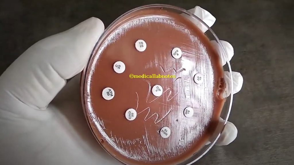
Applying antimicrobial disks on Muller-Hinton agar (MHA) for antimicrobial susceptibility testing (AST)

Measuring Zone of Inhibition (ZOI) for antimicrobial disks
Apply E-Test strip on Muller-Hinton agar (MHA) to determine the MIC
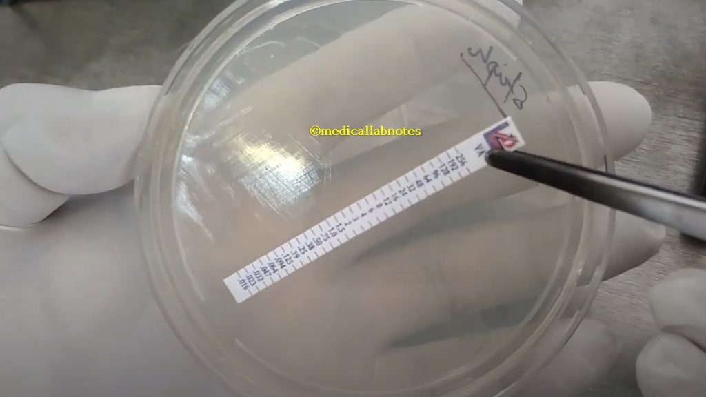
MIC of Vancomycin Determination by E-strip for Staphylococcus aureus

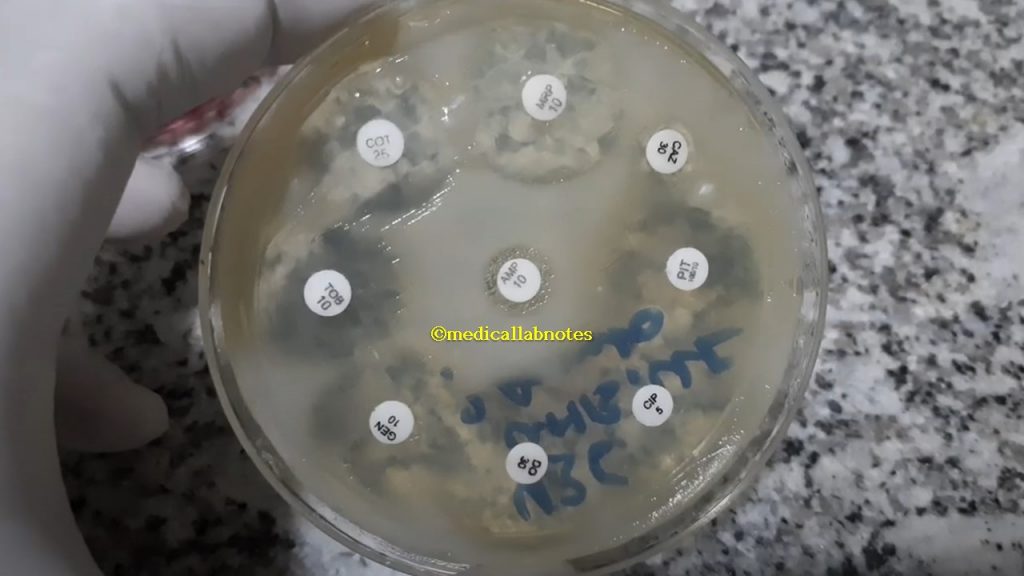
Pseudomonas aeruginosa Antimicrobial Sensitivity Testing (AST) Pattern

Enterobacteriaceae (Escherichia coli) AST pattern
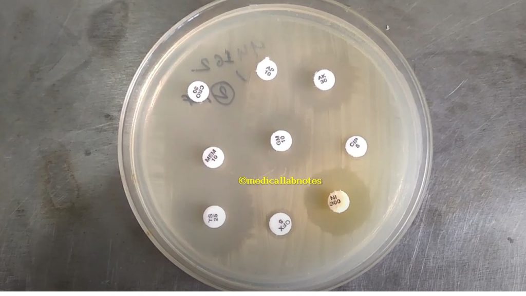
Staphylococcus Antimicrobial susceptibility Testing Pattern

Acinetobacter Antibiogram Pattern
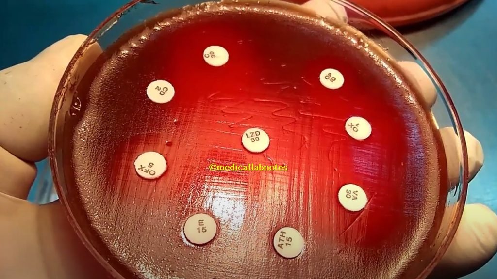
Beta-hemolytic streptococci (BHS) Antimicrobial Susceptibility Testing Pattern

Streptococcus pneumoniae Antibiogram assaying pattern on modified MHA
Haemophilus Antibiogram on modified MHA
Further Readings
- https://labmal.com/product/nutrient-agar-500g/
- http://himedialabs.com/TD/M001.pdf
- https://catalog.hardydiagnostics.com/cp
- https://en.wikipedia.org/wiki/Nutrient_agar
- https://catalog.hardydiagnostics.com/cp_prod/Content/hugo/CRITN-MacConkeyAgar.htm
- https://www.ncbi.nlm.nih.gov/books/NBK557394/
- http://www.himedialabs.com/TD/M081B.pdf
- http://www.oxoid.com/UK/blue/prod_detail/prod_detail.asp?pr=CM0115&cat=&c=UK&lang=EN
- Bailey & Scott’s Diagnostic Microbiology. Editors: Bettey A. Forbes, Daniel F. Sahm & Alice S. Weissfeld, 12th ed 2007, Publisher Elsevier.
- Pelczar M. J. Jr., Reid R. D., Chan E. C. S., 1977, Microbiology, 4th Ed., Tata McGraw-Hill Publishing Company Ltd, New Delhi.
- Clinical Microbiology Procedure Handbook, Chief in editor H.D. Isenberg, Albert Einstein College of Medicine, New York, Publisher ASM (American Society for Microbiology), Washington DC.
- Koneman E. W., Allen S. D., Janda W. M., Schreckenberger P. C., Winn W. C. Jr., 1992, Colour Atlas and Textbook of Diagnostic Microbiology, 4th Ed., J. B. Lippincott Company.
- Spector W. S., (Ed.), 1961, Handbook of Biological Data, W. B. Saunders Company, Philadelphia and London.
- https://www.asm.org/getattachment/7ec0de2b-bb16-4f6e-ba07-2aea25a43e76/protocol-2885.pdf
- Colour Atlas and Textbook of Diagnostic Microbiology. Editors: Koneman E.W., Allen D.D., Dowell V.R. Jr, and Sommers H.M.
- https://www.bd.com/europe/regulatory/Assets/IFU/Difco_BBL/211086.pdf
- Mackie and Mc Cartney Practical Medical Microbiology. Editors: J.G. Colle, A.G. Fraser, B.P. Marmion, A. Simmous, 4th ed, Publisher Churchill Living Stone, New York, Melborne, Sans Franscisco 1996.
- Textbook of Diagnostic Microbiology. Editors: Connie R. Mahon, Donald G. Lehman & George Manuselis, 3rd edition2007, Publisher Elsevier.
- Manual of Clinical Microbiology. Editors: P.R. Murray, E. J. Baron, M. A. Pfaller, F. C. Tenover, and R. H. Yolken, 7th ed 2005, Publisher ASM, USA
- https://www.thermofisher. com/order/catalog/product/R01293#/R01293
- https://www.sciencedirect.com/topics/immunology-and-microbiology/chocolate-agar
- https://jcm.asm.org/content/jcm/20/4/822.full.pdf
- https://www.ncbi.nlm.nih.gov/pmc/articles/PMC533390/
- https://anaerobesystems.com/products/plated-media/chocolate-agar-choc
- https://www.ncbi.nlm.nih.gov/pmc/articles/PMC271442/
- https://www.thermofisher.com/order/catalog/product/R454082#/R454082
- http://himedialabs.com/TD/M173.pdf
- https://www.sigmaaldrich.com/content/dam/sigma-aldrich/docs/Sigma-Aldrich/Datasheet/1/70191dat.pdf
- http://www.tulipgroup.com/MicroExpress/Accumix/PackInsert/Dehydrated%
- https://www.researchgate.net/post/Why_muellerhinton_agar_is_used_in_routine_antibiotic_susceptibility_testing
- http://www.himedialabs.com/HML/images/literature/pdf/100000027/68.pdf
- https://en.wikipedia.org/wiki/Mueller-Hinton_agar
I like reading through an article that will make men and women think. Also, many thanks for allowing me to comment!
Pretty! This has been a really wonderful article. Many thanks for supplying these details.|