
Introduction
Table of Contents
The Mycobacteria Growth Indicator Tube (MGIT) system is a high-throughput, automated method optimized for the rapid isolation and detection of Mycobacterium tuberculosis complex and non-tuberculous mycobacteria (NTM).
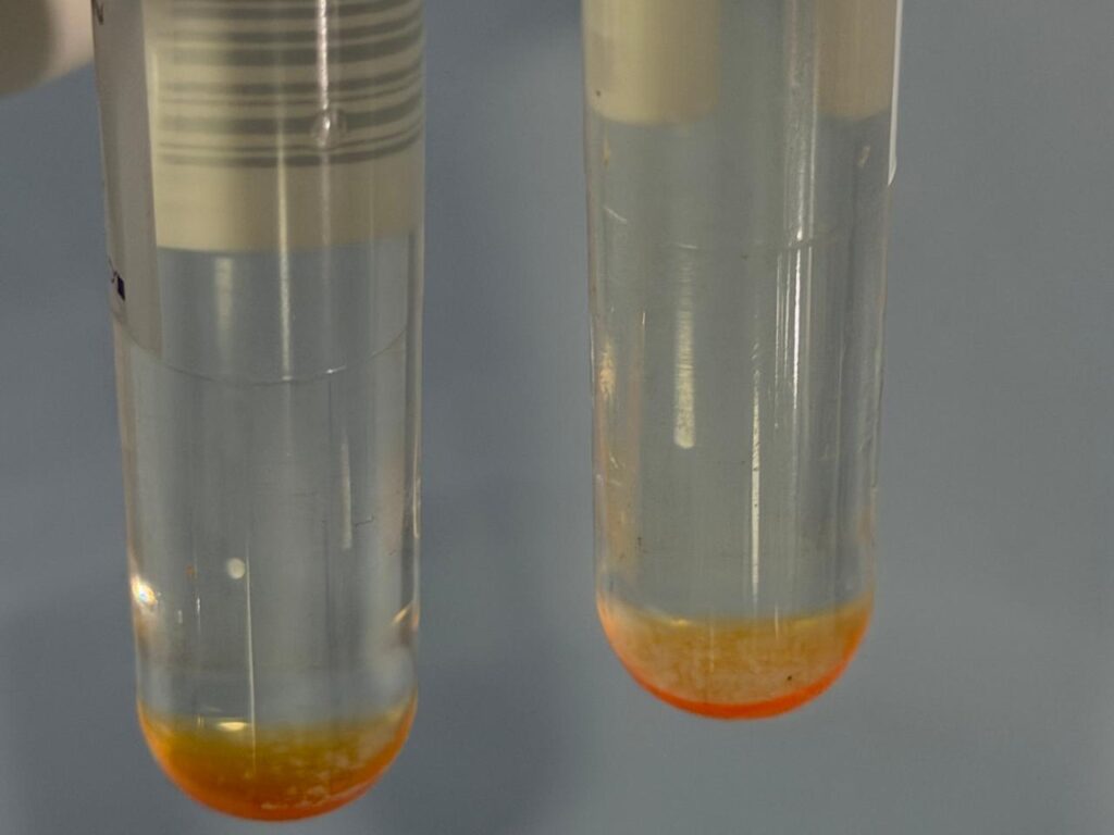
- The Medium: Each tube contains 7.0 ml of modified Middlebrook 7H9 liquid broth, which provides nutrient-rich conditions for faster growth compared to traditional solid media.
- The Principle: The system relies on a fluorescence quenching-based oxygen sensor embedded in silicone at the bottom of the tube.
- The Mechanism: High levels of dissolved oxygen initially quench the sensor’s fluorescence. As actively respiring bacteria consume oxygen, the sensor is unquenched, causing the tube to fluoresce under UV light or inside an automated system like the BD BACTEC MGIT 960 Instrument.
[Initial Stage] Oxygen is high —–> Quenches sensor —–> NO FLUORESCENCE
[Bacterial Growth] Oxygen consumed —-> Frees sensor —–> HIGH FLUORESCENCE (Signal)
Identification and Classification
In standard clinical practice, a processed specimen is inoculated into the tube alongside a growth supplement and an antibiotic mixture called PANTA (Polymyxin B, Amphotericin B, Nalidixic Acid, Trimethoprim, and Azlocillin) to suppress normal flora. Tubes are classified into three distinct categories:
1. MGIT Positive
- Definition: The tube exhibits a high level of fluorescence, prompting the automated instrument to flag it as “positive”.
- Identification: Confirmation requires a mandatory Acid-Fast Bacilli (AFB) smear using Ziehl-Neelsen (ZN) or auramine staining.
- Visual Clues: True positive mycobacterial growth typically shows acid-fast positive bacilli, often forming characteristic cord-like aggregates (cording) under the microscope. Gram staining is performed simultaneously to rule out non-mycobacterial contamination.
2. MGIT Negative
- Definition: The tube fails to flag positive within the standard validation period.
- Identification: The automated instrument officially designates the tube as negative after 42 days of continuous incubation with no significant change in oxygen consumption.
3. MGIT Contamination
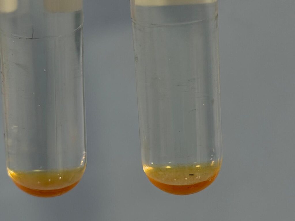
- Definition: Non-target microorganisms (bacteria, yeasts, or molds) overgrow the culture, consuming oxygen and triggering a premature positive signal.
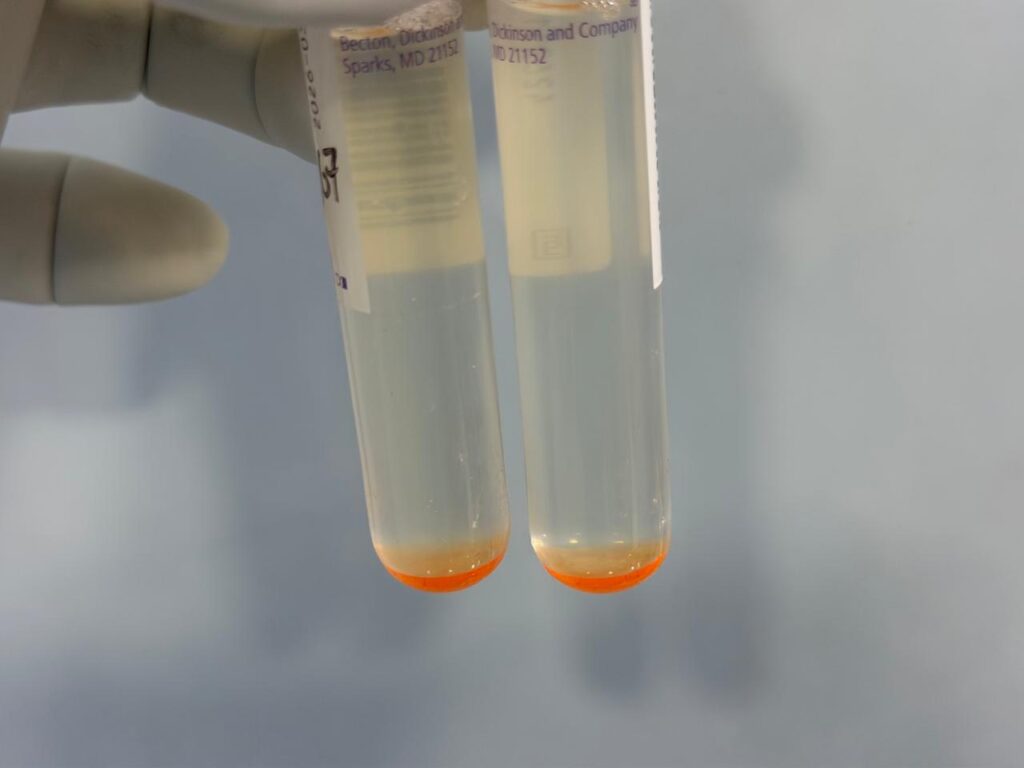
- Identification: When a flagged tube is smeared, it shows acid-fast negative organisms on the ZN stain, or reveals dense Gram-positive/Gram-negative bacteria or fungal elements under a Gram stain. The liquid media may also appear excessively turbid or show a distinct color change.
Possible Reasons for Contamination
Liquid culture media like Middlebrook 7H9 are highly enriched, making them much more susceptible to contamination than solid egg-based slants.
- Survival of Normal Flora: Common respiratory and oral cavity flora—such as Bacillus spp., Staphylococcus spp., Pseudomonas spp., and various yeasts—can survive if the initial chemical decontamination step (like NALC-NaOH) is too mild or brief.
- Transport and Storage Delays: Delays in shipping clinical specimens to the lab, especially in warm climates, allow non-mycobacterial local flora to multiply extensively, overwhelming the decontamination process.
- Inadequate PANTA Potency: Reconstituted PANTA antibiotics may degrade if stored improperly, failing to suppress background bacterial or fungal growth in the tube.
- Environmental/Laboratory Vectors: Contamination can occur during inoculation from unsterilized laboratory equipment, non-sterile water sources, or through cross-contamination via shared reagent dispensers.
Keynotes
- The Golden Rules: A positive signal from the MGIT machine indicates biological respiration, not necessarily tuberculosis. Always verify a positive vial with an AFB smear and a Gram stain to check for mixed growth or pure contamination.
- False Negatives: A small subset of M. tuberculosis strains grow poorly or slowly, occasionally staying under the instrument’s detection threshold and resulting in a false negative at 42 days.
- Salvaging Contaminated Tubes: Do not immediately discard contaminated cultures. Vials containing mixed growth can often be successfully re-decontaminated (e.g., using 4% NaOH or mild oxalic acid) and re-inoculated to recover the target mycobacteria.
- Speed vs. Specificity: While the MGIT system dramatically cuts down the time to detection to a matter of days, rapid downstream assays like lateral-flow MPB64 antigen tests or nucleic acid amplification tests (NAATs) must follow to differentiate true M. tuberculosis complex from non-tuberculous mycobacteria (NTM).
Further Readings
- https://www.finddx.org/wp-content/uploads/2023/02/20061101_rep_mgit_manual_FV_EN.pdf
- https://pmc.ncbi.nlm.nih.gov/articles/PMC130878/
- https://journals.asm.org/doi/pdf/10.1128/jcm.35.8.2068-2071.1997
- https://www.ncbi.nlm.nih.gov/pmc/articles/PMC7383301/
- https://pmc.ncbi.nlm.nih.gov/articles/PMC7383301/
- https://pmc.ncbi.nlm.nih.gov/articles/PMC4963527/
- https://en.wikipedia.org/wiki/Mycobacteria_growth_indicator_tube
- https://onlinelibrary.wiley.com/doi/10.1155/2020/1721020
- https://ajlmonline.org/index.php/ajlm/article/view/537/850
- https://journals.asm.org/doi/10.1128/jcm.02243-10
- https://onlinelibrary.wiley.com/doi/10.1155/2016/5972021
- https://pmc.ncbi.nlm.nih.gov/articles/PMC11557378/
- https://pmc.ncbi.nlm.nih.gov/articles/PMC10321564/
- https://eprints.nirt.res.in/465/1/199808.pdf
- https://pmc.ncbi.nlm.nih.gov/articles/PMC5204086/
- http://whocctblab.fondazionesanraffaele.it/uploads/2/0/8/2/20828554/ios_ebp_dma_007_identification_of_m.tb_rev_1.pdf