
Introduction
Table of Contents
The BD BACTEC™ MGIT™ 960 (Mycobacteria Growth Indicator Tube) is a fully automated, high-volume culture system designed for the rapid detection of mycobacteria and subsequent drug susceptibility testing (DST). Developed by Becton Dickinson (BD), this nonradiometric system addresses the critical clinical need for accelerated tuberculosis (TB) diagnostics.
Traditional solid culture media, such as Löwenstein-Jensen (LJ) slants, can take 4 to 8 weeks to yield visible colonies. The MGIT™ 960 continuously monitors liquid culture tubes, reducing detection time to days or weeks. Housing up to 960 tubes simultaneously, it serves as a cornerstone instrument in high-throughput microbiology and reference laboratories globally.
Principle
The system operates on a fluorometric sensor technology that detects bacterial respiration rather than visible colony formation.
Chemical Components
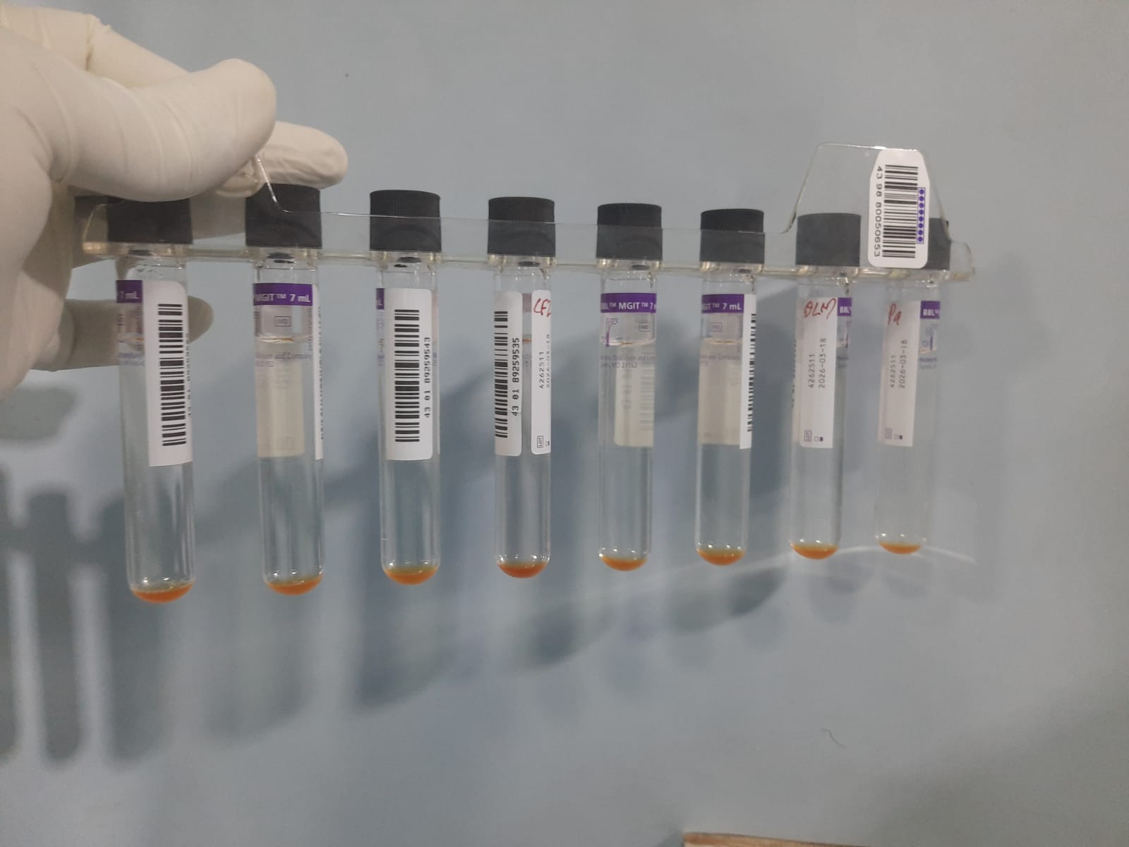
- The Medium: Each tube contains 7.0 mL of modified Middlebrook 7H9 liquid broth, optimized to support the rapid growth of mycobacteria.
- The Sensor: A fluorescent compound (an oxygen-quenched fluorochrome) is embedded in a layer of silicone at the bottom of each tube.
Biological Principle
- Oxygen Quenching: In an uninoculated or negative tube, dissolved oxygen is abundant in the liquid broth. This abundant oxygen continuously quenches (represses) the fluorescence of the sensor.
- Depletion by Respiration: As mycobacteria multiply, they consume the dissolved oxygen within the sealed environment of the tube.
- Fluorescence Activation: Once the oxygen level drops below a specific threshold, the quenching effect ceases. The fluorochrome becomes uninhibited and begins to emit free fluorescence when exposed to blue light.
Working Mechanism
The operational workflow from sample collection to raw machine readout involves strict technical steps:
[Sample Collection] → [Decontamination & Digestion (NALC-NaOH)] →[Reagent Addition (OADC Enrichment, particularly in case of DST case otherwsie only PANTA Antibiotic Blend)] → [Inoculation into MGIT Tube & Instrument Loading] → [Automated Hourly Photodetector Scanning] → [Machine Log: Growth Units (GU) Calculation]
Step 1: Specimen Preparation & Decontamination
Raw clinical specimens (like sputum or gastric washings) naturally contain fast-growing oral flora that will outgrow slow-growing mycobacteria. Samples must undergo decontamination and digestion, typically using the NALC-NaOH (N-acetyl-L-cysteine-sodium hydroxide) method, followed by centrifugation to concentrate the bacteria.
Step 2: Tube Enrichment & Suppression
Before adding the processed specimen, two vital reagents are pipetted into the MGIT tube:
- MGIT OADC Enrichment: Provides essential nutrients (oleic acid, albumin, dextrose, and catalase) to stimulate mycobacterial metabolic pathways.
- MGIT PANTA: A lyophilized antibiotic cocktail (Polymyxin B, Amphotericin B, Nalidixic acid, Trimethoprim, and Azlocillin) that suppresses residual non-mycobacterial bacteria and fungi surviving decontamination.
Step 3: Inoculation and Loading
- Exactly 0.5 mL of the decontaminated, concentrated specimen sediment is inoculated into the prepared MGIT tube.
- The tube is barcode-scanned and loaded into one of the internal instrument racks.
Step 4: Continuous Automated Monitoring
- The instrument maintains an internal incubation temperature of 37°C ± 1°C.
- Every 60 minutes, the instrument’s internal rows of light-emitting diodes (LEDs) flash blue light at the base of each tube.
- Onboard photodetectors read the intensity of the emitted fluorescence.
- The internal software converts this light intensity into mathematical Growth Units (GU).
Result Interpretation
The MGIT™ 960 automatically interprets the fluorescence dynamics over a standard incubation period of up to 42 days (6 weeks) for diagnostic screening.
Automated Instrument Indicators
- Positive Instrument Flag: When the rate of fluorescence increase satisfies the system algorithm (typically crossing a raw threshold of 75 to 100 Growth Units within a set timeframe), a green indicator light on the corresponding drawer flashes, and the instrument logs the tube as positive.
- Negative Instrument Flag: If a tube shows no significant rise in GU by the end of the 42-day testing period, the instrument registers the tube as negative, indicating no viable mycobacteria were detected.
Mandatory Manual Follow-up for Positive Tubes
Because the oxygen sensor responds to respiration from any living microorganism, an instrument-positive flag does not guarantee the presence of Mycobacterium tuberculosis. Laboratories must execute the following confirmatory cascade immediately upon a positive alert:
| Confirmatory Test | Purpose | Action Based on Result |
| Acid-Fast Bacilli (AFB) Smear | Verifies morphological presence of mycobacteria. | If positive, confirms mycobacteria. If negative, suggests contamination or low bacterial load. |
| Blood Agar/Chocolate Agar Culture | Checks for non-mycobacterial contamination. | Growth within 24–48 hours indicates contamination; requires re-decontamination of the tube. |
| Rapid Antigen/Molecular Testing | Differentiates M. tuberculosis complex from NTMs. | Positive MPT64 antigen test or PCR confirms M. tuberculosis complex. |
Clinical Applications
Detection of Acid-Fast Bacilli (AFB)
The primary diagnostic application is isolating viable mycobacteria from clinical specimens. This includes pulmonary samples (sputum, bronchoalveolar lavage) and extrapulmonary samples (cerebrospinal fluid, tissue biopsies, pleural fluids).
Differentiation of Mycobacterial Species
While it does not identify species on its own, the harvested liquid biomass from a positive MGIT tube provides high-yield starter material for rapid immunochromatographic assays (like MPT64 antigen detection) or line probe assays to distinguish Mycobacterium tuberculosis complex (MTBC) from Non-Tuberculous Mycobacteria (NTM).
Drug Susceptibility Testing (DST)
The system is widely validated for first-line and second-line Drug Susceptibility Testing.
- Methodology: The system compares the growth rate of a patient’s isolate in a drug-containing MGIT tube against a drug-free control tube (diluted 1:100 to represent a 1% critical population). [1, 2]
- First-line DST: Evaluates susceptibility to Streptomycin, Isoniazid, Rifampicin, and Ethambutol (SIRE), as well as Pyrazinamide (PZA), which requires a dedicated, low-pH MGIT medium to match drug activation requirements.
- Second-line DST: Conducts testing for fluoroquinolones (e.g., Ofloxacin, Moxifloxacin) and injectable agents (e.g., Amikacin, Capreomycin) to map Multi-Drug Resistant (MDR) and Extensively Drug-Resistant (XDR) patterns.
Keynotes
- Turnaround Time (TAT) Efficiency: Average detection time for M. tuberculosis falls to 10–14 days compared to the 28–42 days required by solid media cultures.
- Nonradiometric and Needleless Safety: Replaces older BACTEC™ 460 systems that relied on carbon-14 (¹⁴C) radioactive substrates and needle aspiration. The MGIT™ 960 relies entirely on optical scanning through the intact outer plastic tube, minimizing biohazard exposure.
- Contamination Sensitivity: The rich Middlebrook 7H9 liquid matrix is sensitive to breakthrough contamination. If the PANTA antibiotic cocktail fails or the NALC-NaOH decontamination step is under-dosed, rapid-growing contaminants (e.g., Pseudomonas, Bacillus, or fungi) will consume the oxygen, triggering false-positive system alerts for TB.
- Biosafety Mandate: Due to the liquid propagation of high-concentration airborne pathogens, all inoculation, opening of positive MGIT tubes, and subsequent testing must occur strictly within a certified Biosafety Level 3 (BSL-3) facility using Class II Biosafety Cabinets.
Further Readings
- https://www.cdc.gov/tb/hcp/testing-diagnosis/clinical-and-laboratory-diagnosis.html
- https://ntep.in/node/1547/CP-specimen-preparation-lc-and-lpa-labs
- https://ntblcp.org.ng/content/uploads/2023/06/National-SOP-for-TB-laboratory-diagnosis-Jan-2017.pdf
- https://pmc.ncbi.nlm.nih.gov/articles/PMC11434331/
- http://whocctblab.fondazionesanraffaele.it/uploads/2/0/8/2/20828554/ios_ebp_dma_004_specimen_processing_for_culture_rev_1.pdf
- https://www.ntp.gov.bd/wp-content/uploads/2024/06/SOP_MTB-Culture_DST.pdf
- https://pmc.ncbi.nlm.nih.gov/articles/PMC3108717/
- https://www.moleculartb.org/files/documents/10
- https://hardydiagnostics.com/media/assets/product/documents/DecontamRgntRecovMyco.pdf
- https://www.alphatecsystems.com/files/dfu/L003455.F%20-%20Sodium%20Hydroxide%20Solution,%204%25%20%20Sodium%20Citrate%20Solution,%202.94%25.pdf
- https://www.ntep.in/node/1466/CP-culture-specimen-processing-advantages-and-disadvantages-nalc-naoh-method
- https://www.sciencedirect.com/science/article/abs/pii/S0732889313003763
- https://pmc.ncbi.nlm.nih.gov/articles/PMC139704/
- http://whocctblab.fondazionesanraffaele.it/uploads/2/0/8/2/20828554/_ios_ebp-specimen_processing_for_culture.pdf