
Introduction
Table of Contents
KOH mount (Potassium Hydroxide preparation) is a rapid, simple, and cost-effective microscopic technique used to detect fungal elements directly in clinical specimens.
When applied to Bronchoalveolar Lavage (BAL) samples, it is an essential diagnostic tool for identifying pulmonary fungal infections, especially in the ICU, oncology, transplant, and immunocompromised patients.

KOH dissolves mucus, cellular debris, and proteins present in the BAL fluid, clearing the background while
, preserving the chitin-rich fungal cell walls, making fungal structures easily visible under the microscope.

BAL KOH mount is extremely helpful in diagnosing invasive fungal infections, opportunistic mycoses, and mixed fungal–bacterial infections in the lungs.
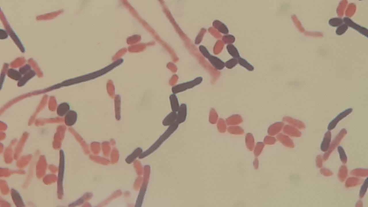
Fungal Elements Observed in BAL KOH Mount

1. Yeast Cells
- Round/oval, refractile structures
- May show budding (blastoconidia)
- Common species: Candida, Cryptococcus
- Cryptococcus may show a halo-like capsule clearing
2. Pseudohyphae
- Elongated, sausage-like, with constrictions at septa
- Seen commonly in Candida albicans
- Indicates active infection rather than colonization
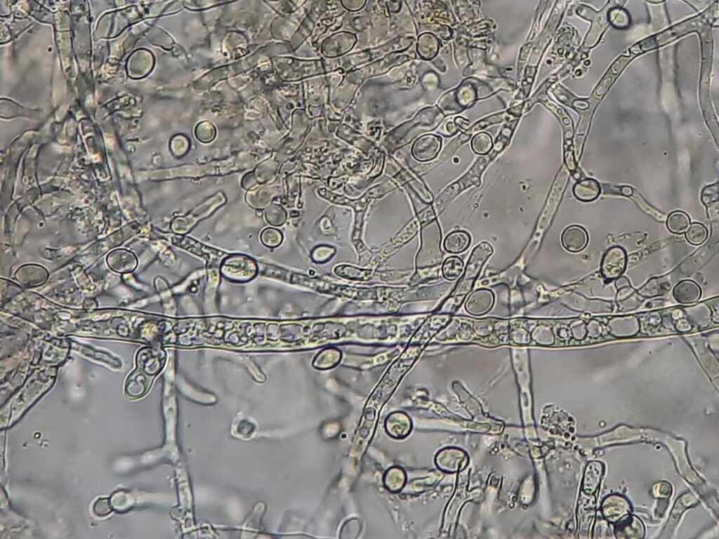
3. True Hyphae
- Long, filamentous, branched structures
- Common in Aspergillus, Fusarium, Mucorales (broad, non-septate)
- Branching patterns are diagnostic clues:
- Aspergillus → thin, septate, acute angle branching
- Mucorales → broad, ribbon-like, non-septate hyphae
4. Conidia / Spores
- Small, round refractile bodies
- Found in Aspergillus, Penicillium, and other molds
5. Pneumocystis jirovecii Structures (Occasional)
- Foamy alveolar casts
- Crushed “ping-pong ball” cysts
- Better visualized with GMS or Giemsa, but KOH may show cyst-like bodies
Applications of Fungal Elements in KOH Mount of BAL (Bronchoalveolar Lavage) Microscopy
- Rapid Screening Test: Provides immediate detection of fungal elements before culture or molecular tests.
- Diagnosis of Pulmonary Fungal Infections: Useful in identifying invasive aspergillosis, mucormycosis, pneumocystis pneumonia, and Candida pneumonia.
- Critical Care and Immunocompromised Patients: Essential for diagnosing fungal infections in cancer, HIV/AIDS, transplant, and ICU patients.
- Guiding Antifungal Therapy: Helps clinicians initiate early antifungal treatment, especially in emergency settings.
- Corroborating Culture and Molecular Findings: Microscopy supports results from culture, PCR, sequencing, or antigen tests (e.g., galactomannan, β-D-glucan).
Keynotes on Fungal Elements in KOH Mount of BAL

- KOH concentration used: 10–20%
- Fungal elements appear refractile, clear, and well-defined against a cleared background
- BAL KOH mount is rapid but not species-specific—confirm with culture or molecular tests
- Helpful for differentiating colonization versus invasive infection when correlated clinically
- Essential for diagnosing fungal pneumonia in oncology, ICU, and immunocompromised patients
- Should be interpreted by an experienced microbiologist to avoid artifacts (e.g., mucus threads, crystals)
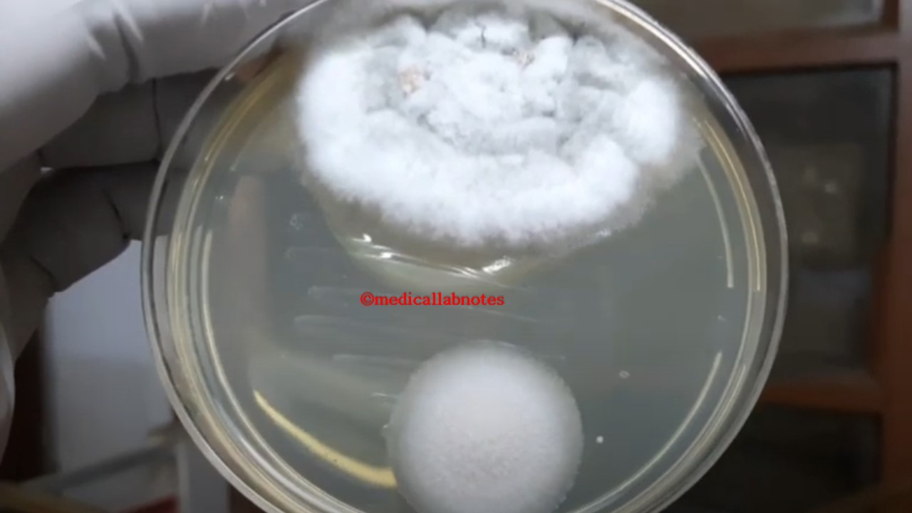
- Always pair with SDA culture, GMS stain, PAS stain, or PCR for confirmation

Further Readings
- https://pmc.ncbi.nlm.nih.gov/articles/PMC10492593/
- https://www.researchgate.net/figure/KOH-mount-of-bronchoalveolar-lavage-fluid-obtained-after-bronchoscopy-Magnification-3440_fig3_23655567
- https://pmc.ncbi.nlm.nih.gov/articles/PMC10492593/#:~:text=On%20the%20other%20hand%2C%20many,to%20provide%20more%20appropriate%20treatment.
- https://www.researchgate.net/figure/KOH-mount-of-bronchoalveolar-lavage-fluid-obtained-after-bronchoscopy-Magnification-3440_fig3_23655567
- https://www.1mg.com/labs/test/koh-staining-sputum-35070
- https://www.dalynn.com/dyn/ck_assets/files/tech/RP85-25.pdf
- https://drkothiwalaskineva.com/medical-dermatology/koh-mount-for-fungal-diseases/
- https://ijdvl.com/skin-scraping-and-a-potassium-hydroxide-mount/
- https://ritm.gov.ph/laboratory-services/fungal-culture-and-susceptibility-test-urineinclusion1-potassium-hydroxide-mount-koh-29/
- https://en.wikipedia.org/wiki/KOH_test#:~:text=Collection:%20Skin%2C%20nail%2C%20or,follicles%20from%20the%20infected%20site.
- https://www.ncbi.nlm.nih.gov/books/NBK430762/