Isolation and Preliminary Identification of Bacterial and Yeast Colonies on Sabouraud Dextrose Agar Using the Wet Mount Technique

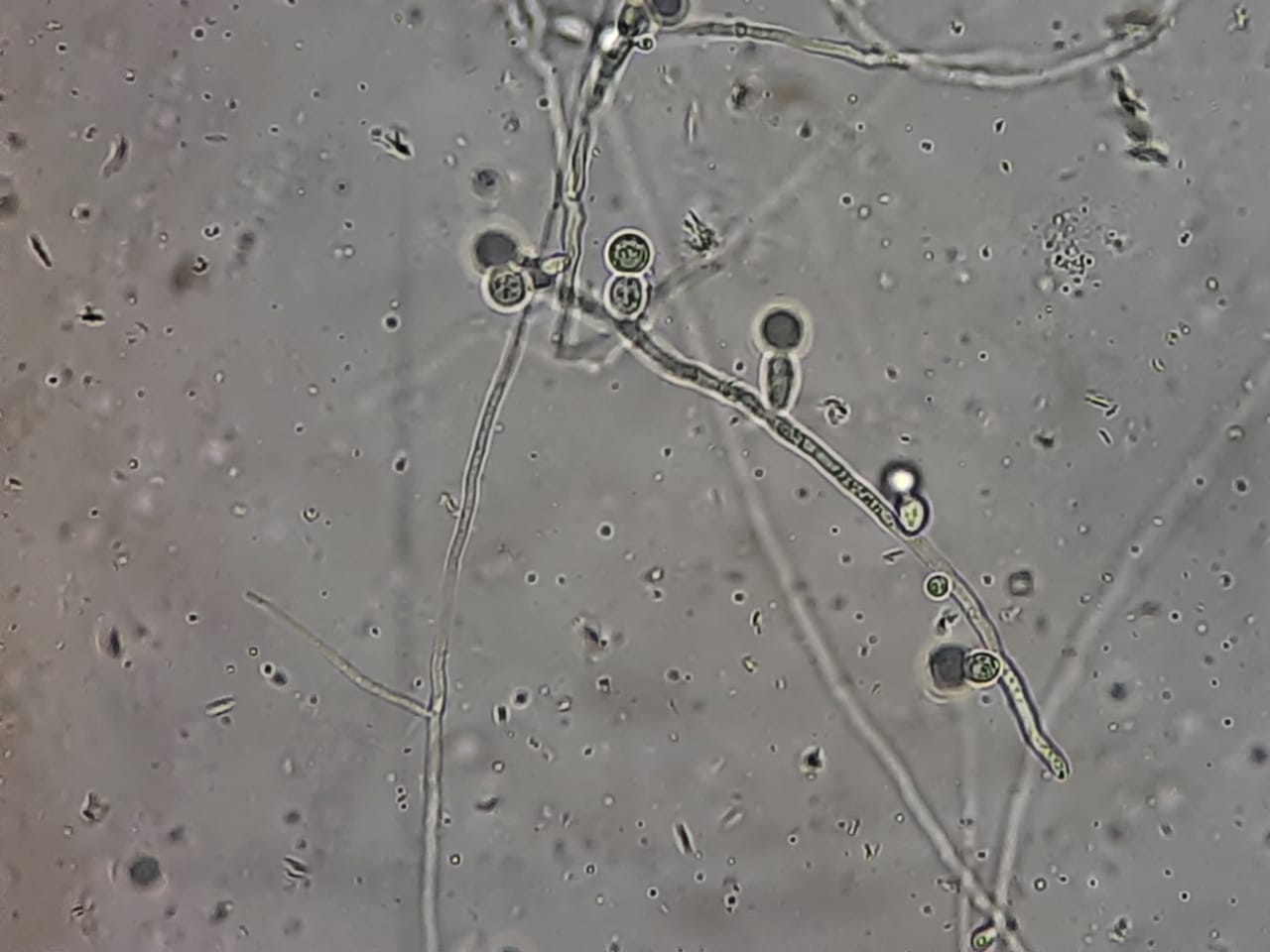
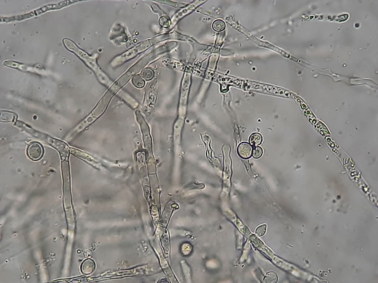
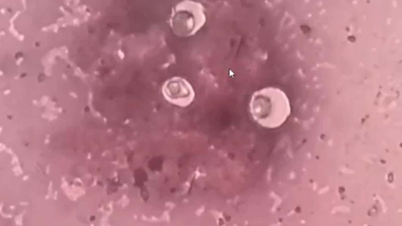
Introduction Sabouraud Dextrose Agar (SDA) is a commonly used culture medium in clinical mycology laboratories for the isolation of fungi, particularly yeasts and molds. Due to its acidic pH and high dextrose concentration, SDA favors fungal growth; however, bacteria and yeasts may also grow, especially …