Fungal Elements in KOH Mount of Sputum Microscopy-Introduction, Fungal Elements Observed in Sputum KOH Mount, Applications, and Keynotes

Fungal Elements in KOH Mount of Sputum Microscopy at a magnification of 1600X

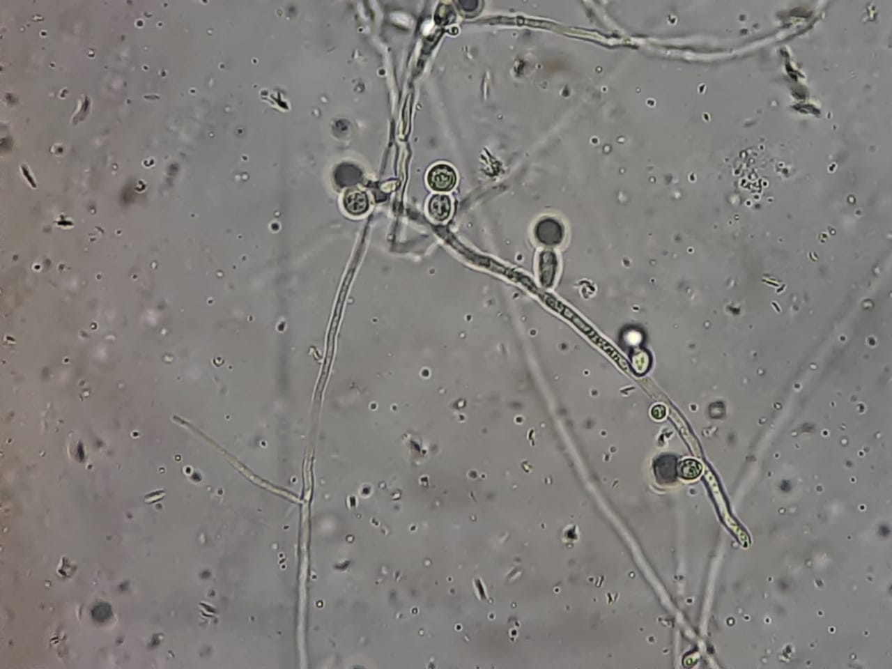
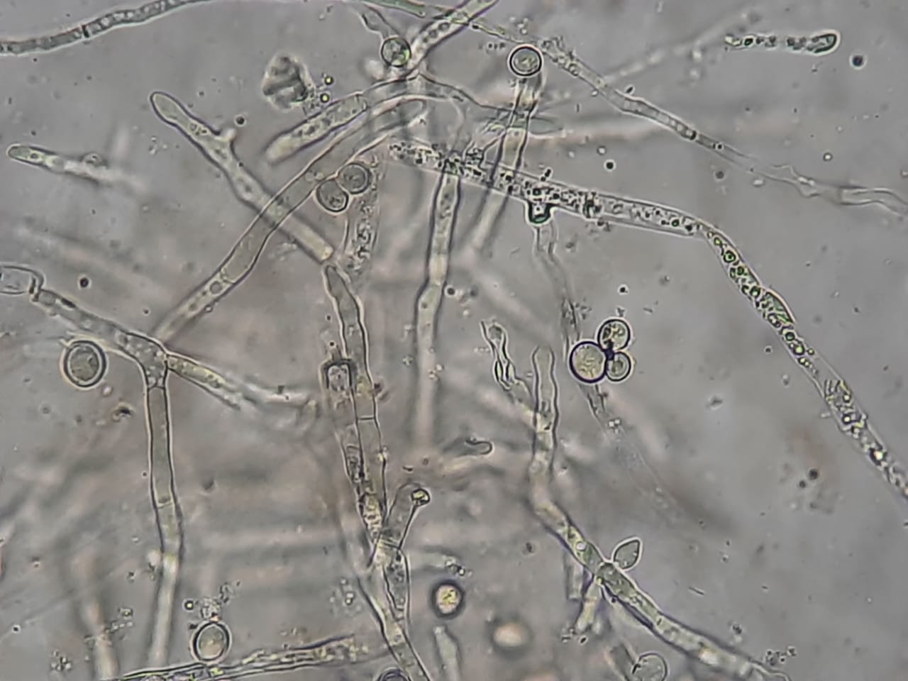
Introduction KOH mount of sputum is a rapid, direct microscopic technique used to detect fungal elements in the respiratory tract. A 10–20% Potassium Hydroxide (KOH) solution digests mucus, epithelial cells, and debris in sputum, while preserving the chitin-rich fungal cell walls, making them appear clear, …

Read more

Fungal Elements in KOH Mount of BAL (Bronchoalveolar Lavage) Microscopy- Introduction, Fungal Elements Observed in BAL KOH Mount, Applications, and Keynotes

Fungal Elements in wet mount of culture of BAL specimen

Introduction KOH mount (Potassium Hydroxide preparation) is a rapid, simple, and cost-effective microscopic technique used to detect fungal elements directly in clinical specimens.When applied to Bronchoalveolar Lavage (BAL) samples, it is an essential diagnostic tool for identifying pulmonary fungal infections, especially in the ICU, oncology, …

Read more

Fungal Elements in KOH Mount of Urine Microscopy: Introduction, Fungal Elements Observed in Urine KOH Mount, Applications, and Keynotes

Fungal elements in KOH Mount of Urine Microscopy- Introduction, Fungal Elements Observed in Urine KOH Mount, Applications, and Keynotes

Introduction Potassium Hydroxide (KOH) mount is a simple, rapid, and cost-effective microscopic technique used to detect fungal elements in various clinical specimens, including urine samples. When fungal infection of the urinary tract (funguria) is suspected—especially in immunocompromised, diabetic, or catheterized patients—KOH mount helps visualize yeast …

Read more

Comparative Microscopy of Common Clinical Fungi-Introduction, Table, and Appearance at Different Magnifications

Aspergillus in LPCB tease mount of culture

Introduction Microscopy remains one of the cornerstones of clinical mycology, providing a rapid and inexpensive method for the direct detection and preliminary identification of fungi. Comparative microscopy is particularly valuable in distinguishing the morphological features of common clinical fungi, many of which exhibit overlapping clinical …

Read more

Criteria and Decision Framework for Differentiating True Infection from Colonization or Contamination by Aspergillus niger in Cancer Patients

Aspergillus niger growth on SDA

Evaluate the Clinical Context Sample Collection & Repeatability Microscopy & Direct Detection Quantitative Culture & Sterility of Site Serological & Molecular Evidence Radiological Correlation Practical Interpretation Framework Finding Likely Contaminant Possible Colonizer Likely Pathogen Single isolate from 1 sample ✅ ✅ ❌ Repeated isolation from …

Read more

Yeasts vs Molds: Detailed Comparison, Mixed Growth Isolation, Approach to Mixed Growth Handling, and Mixed Growth Interpretation

Yeast and mold mixed growth on SDA

Yeasts vs Molds: Detailed Comparison Feature Yeasts Molds Definition Unicellular fungi Multicellular filamentous fungi Structure Oval/round, single cells Hyphae form mycelium; septate or aseptate Reproduction Asexual (budding or fission); some sexual forms Asexual (spores like conidia/sporangia); sexual spores Hyphae Absent, but pseudohyphae may be present …

Read more

Funguria-Introduction, Common fungi, Clinical Feature, Lab Diagnosis, Treatment, and Prevention

Urine sample for fungal culture and KOH mount

Introduction Funguria refers to the presence of fungi in the urine, either due to colonization, contamination, or true urinary tract infection (UTI). It is increasingly reported in hospitalized or catheterized patients, particularly those with immunosuppression, antibiotic overuse, or diabetes. Most cases are asymptomatic, but it …

Read more